Leiomyoma of the Trachea: A Rare Tracheal Tumor
Iftekhar Ahmed1 ,Niaz Hussain Soomro 2 , Muhammad Ashhal Iftekhar3,, Sana Siddiqui4
1Ojha Institute of Chest Diseases, Dow University of Health Sciences, Karachi
Abstract
Background:Primary tumors of the trachea are rare and among these 1% are leiomyomas. We report a case of a tracheal leiomyoma diagnosed incidentally in a 45-year-old male farmer who presented with recurrent dyspnea for four years. He was initially misdiagnosed as asthma and treated with a combined approach. The patient however experienced worsening symptoms, including cough, hemoptysis, weight loss, and respiratory failure. Imaging and bronchoscopy revealed a tracheal mass, later diagnosed as leiomyoma. The patient underwent tumor resection via rigid bronchoscopy, resulting in successful removal and tracheal recanalization. Postoperatively, the patient showed improvement, and follow-up examinations confirmed resolution of symptoms.
Key words: Leiomyoma, Tracheal Tumor, Tracheal Mass.
Primary tracheal tumors are rare, and 90% are malignant.1 Leiomyomas constitute less than 1% of tracheal masses. These benign tumors arise from the smooth muscle cells of the membranous portion of the trachea and have the potential to cause tracheal obstruction. Patients typically present with dyspnea, wheezing, cough, and hemoptysis, and asthma or bronchitis. Given the rarity of these lesions, optimal management has not been defined. Broncohscopic local surgical excision and partial tracheal resection have all been described. One report of recurrence after resection has been published. The incidence of recurrence following local excision is unknown.
Case Report: A 45-year-old male patient, farmer by profession and a non-smoker, presented with a previous history of recurrent episodes of dyspnea for the past 4 years. He was misdiagnosed with Asthma and was being treated for the same with bronchodilators and inhalation corticosteroids and was unresponsive to treatment. He currently presented a three-month history of dry cough, hemoptysis, worsening exertional dyspnea and significant weight loss (approximately 15 kg). His CT scan done in the previous hospital revealed a mass at the trachea above the carina. Upon admission he was dyspneic, tachypneic (36 breaths/min) and maintaining 80% oxygen saturation via 10 Liter 02 having 110bpm pulse rate. Chest auscultation revealed wheezing bilaterally. Arterial blood gases revealed Type 1 respiratory failure. Sputum studies for tuberculosis and routine culture were negative. Other routine laboratory investigations were within normal limits.
Detailed investigations were done upon previous hospitalization which included a CT scan with contrast and a fiberoptic bronchoscopy. The CT scan showed a mass of 3.1*2.9cm in the trachea just above the carina. Fiber optic bronchoscopy showed polypoidal growth at distal trachea, however, unfortunately the biopsy taken at that time was inconclusive. A follow-up flexible bronchoscopy at our institution was taken which showed a growth in the distal trachea, therefore the primary carina could not be visualized. There was an acute bleeding from the tracheal growth as well as a drop in the oxygen saturated which resulted in halting the procedure.
It was decided that the patient is a candidate of tumor resection via rigid bronchoscopy. Patient education and counseling was carried out simultaneously regarding risks and benefits of the procedure. After a written consent signed by the patient and family, the patient was anaesthetized by 4% Xylocaine gargles, Propofol and Isoflurane inhalation attached with ventilating rigid bronchoscopy. Rigid bronchoscopy and debulking of the tumor were performed using forceps. The majority of the tumor was successfully removed and trachea was recanalized. Hemostasis secured by Adrenaline-soaked peanuts. Post operatively patient was kept in ICU, he was maintaining saturation of 2 liters of o2, his o2 demand decreased from 10 liters to 2 liters on day 1 and day 2. Later on, he was maintaining oxygen saturation on room air. He was transferred to ward on day 4 and discharged from hospital on day 11. Histopathological results were consistent with leiomyoma. Before discharge flexible bronchoscopy was done to check patency of airway for any residual tumor clot.
Patient was called for follow-up in OPD initially on weekly basis for 4 weeks than monthly for three months. After three months CT scan chest and flexible bronchoscopy was done, both were normal and patient was symptom free.
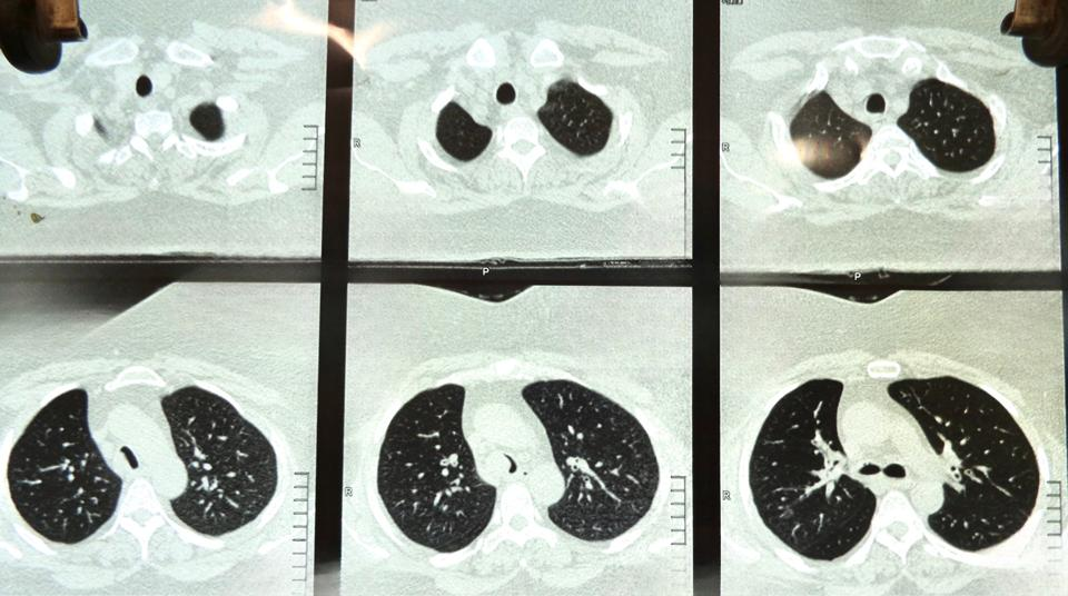
Figure 1: CT scan chest shows- Tumor in the lower trachea. Visually upper trachea and carina are normal.
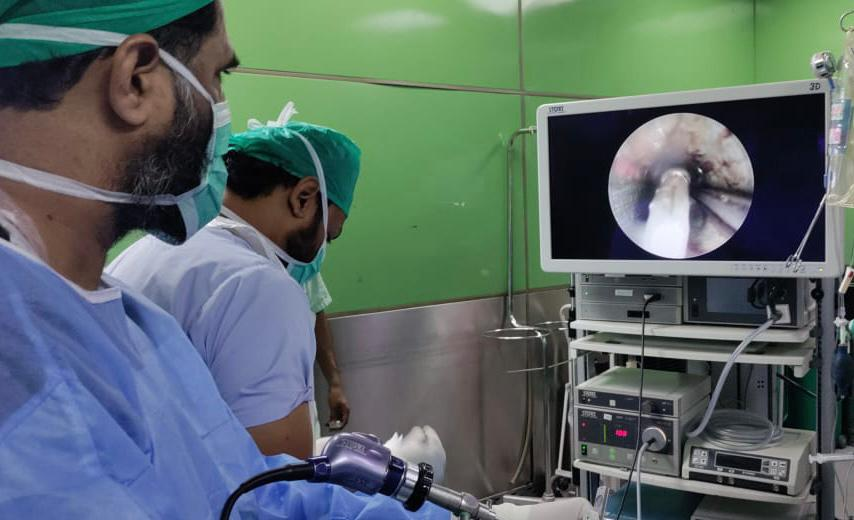
Figure 2: Rigid bronchoscopy being done in the O.R.
Primary tracheal tumors are rare occurrences. Leiomyoma of the trachea is among the scarcer tracheal tumors, accounting for less than 1% of primary tracheal lesions.1 The first reported case of leiomyoma of the trachea was documented in 1955.2 Malignant tumor lesions account for over 90% of tracheal tumors.2 These tumors originate from smooth muscle cells of the membranous trachea. Owing to their location, these tumors have the potential to cause critical airway obstruction.3 Patients will typically present with exertional dyspnea, cough, hemoptysis, wheeze or even asphyxia due to upper airway obstruction. Symptoms are influenced by the size of the tumor and its location.4 Because the symptoms resemble bronchial asthma or bronchitis, these patients are misdiagnosed and often not investigated further.1,4
Given the rarity of this tumor, optimal treatment strategies are yet to be defined.1 Various method for treatment have been described in the literature. There have been a few case reports and case series describing the different management strategies for patients suffering from these tumors.1-6 Various approaches include the classical, such as tracheal resection and local surgical reconstruction and more conservative endoscopic techniques, using a rigid bronchoscope and diverse procedures for bronchoscopic resection such as via forceps, snare excision, cryotherapy, electrocautery, and YAG-laser.5-7 It has been suggested that broad-based tumors should be treated with tracheal resection, as it appears that there is an increased risk of tumor tissue being left behind when endoscopic techniques are employed in such cases, possibly leading to tumor recurrence.5.8 Broncoscopic techniques have been advocated as safe and effective techniques for successful and complete tumor removal.9
An added but crucial aspect of managing these patients is the challenging of anesthesia.
The location of this tumor runs the additional threat of critical airway compromise during procedure, due to bleeding intra-operatively, and possible dislodge of tumor, barring distal airway ventilation. Numerous airway management practices include ventilation through rigid bronchoscope, high-frequency jet ventilation, tracheostomy, cardiopulmonary bypass, or extracorporeal membrane oxygenation.10 Recurrence rates of this tumor have not been established,1 but it has been seen that incomplete bronchoscopic resection in the case of a wide based tumor has resulted in recurrence, which requires surgery and carinal reconstruction.8 The authors therefore agree that while bronchoscopic techniques have provided an effective means of tumor removal, it would be preferred to go to the surgical route for wide-based tumors, to decrease the chances of recurrence.
- Bharadwaj SC, Unruh HW. Leiomyoma of the trachea. The Annals of thoracic surgery. 2012;93(2):669-70. https://doi.org/10.1016/j.athoracsur.2011.07.022
- Dorenbusch AA. Leiomyoma of the trachea; a case report. AMA Arch Otolaryngol. 1955;61(4):470-1. doi:10.1001/archotol.1955.00720020486015
- Garg R, Saini S, Kumar V, Gupta N. Emergency airway management of intratracheal tumor in a patient with respiratory distress. Journal of anaesthesiology, clinical pharmacology. 2017;33(1):133-4. DOI: 10.4103/0970-9185.1682584.
- Koul A. and Sood J. Leiomyoma of the trachea: An anesthetic challange. 2013. Jul-Aug; 57(4): 412–414. doi: 10.4103/0019-5049.118543
- Park JS, Lee M, Kim HK, Choi YS, Kim K, Kim J, et al. Primary leiomyoma of the trachea, bronchus, and pulmonary parenchyma — a single-institutional experience. European Journal of Cardio-Thoracic Surgery. 2011;41(1):41-45. https://doi.org/10.1016/j.ejcts.2011.03.051
- Madan K, Dhooria S, Sehgal IS, Mohan A, Mehta R, Pattabhiraman V, et al. A multicenter experience with the placement of self-expanding metallic tracheobronchial Y stents. 2016;23(1):29-38. DOI: https://doi.org/10.1097/LBR.0000000000000250
- Kitada M, Yasuda S, Ishibashi K, Hayashi S, Matuda Y, Ohsaki Y, et al. Leiomyoma of the Trachea: a case report. Journal of Cardiothoracic Surgery. 2015;10(1):78. DOI 10.1186/s13019-015-0283-0
- 10.hiang Y-Y, Ke C-C, Wu RS-C, Chen K-B, Shen M-L, Poon K-SJIJoG. Endoscopic resection of tracheal tumor in an elderly woman under extracorporeal membrane oxygenation. 2011;5(1):56-8. https://doi.org/10.1016/j.ijge.2011.01.011

An Official Publication of
Islamabad Medical & Dental College
Volume 12 Issue 3
Editorial
Address of Correspondence
Niaz Hussain Soomro
Email:
niaz.hussain@duhs.edu.pk
Cite this article.Ahmed I, Soomro NH, Iftekhar MA, Siddiqui S. Leiomyoma of the Trachea: A Rare Tracheal Tumor.J Islamabad Med Dental Coll. 2023; 11(3): 228-231. DOI: https://doi.org/10.35787/jimdc.v12i3.1021