A Comparison of Functional Outcome of Open Reduction Internal Fixation Versus Ilizarov External Fixation in Complex Proximal Tibial Fractures: Randomized Controlled Trial
Muhammad Farhan Farhat 1 ,Zeeshan Khan Nazim 2Mohammad Osama1Hassaan Ahmad, Bilal Razzaq1
1Department of Orthopaedic Surgery, Pakistan Institute of Medical Sciences (PIMS), Islamabad, Pakistan
1Department of Orthopaedics, Trauma and Spine Surgery, Hayatabad Medical Complex, Peshawar, Pakistan
Objective: This study aimed to compare the functional outcome of Open reduction internal fixation and Ilizarov external fixation in the treatment of complicated proximal tibial fractures
Methodology:This study included 104 patients with Schatzker's grade IV–VI fractures who presented to the orthopedic department at PIMS, Islamabad. Using the lottery approach, the patients were split into two treatment groups at random. Group B uses the Ilizarov External Fixation procedure, while Group A uses the Open Reduction and Internal Fixation (ORIF) technique. Every three weeks, post-operative follow-ups were conducted. Up until the fracture healed, post-operative radiographs were taken every three weeks. Every patient had a 24-week clinical follow-up. Using SPSS v25.0, all of the data were input and processed. The surgery time, VAS score, and length of hospital stay were compared between the two groups using an independent sample t-test. The functional outcome at six months was compared between the two groups using the Chi-square test.
Results:There were 31 (59.6%) men and 21 (40.4%) women in group A, and 36 (69.2%) men and 16 (30.8%) women in group B. In group A, the mean age was 44.87±14.15 years, while in group B, it was 44.25±15.68 years. In terms of functional outcome, 22 (42.3%) of the ORIF technique group had excellent outcomes, while 10 (19.2%), 16 (30.8%), and 4 (7.7%) had good, fair, and poor outcomes, respectively. In contrast, 35 (67.3%) of the Ilizarov technique group had excellent outcomes, while 10 (19.2%), 5 (9.6%), and 2 (3.8%) had good, fair, and poor outcomes, respectively. These results have a statistically significant p-value of 0.024.
Conclusion:Compared to open reduction and internal fixation, Ilizarov external fixation is a more reliable method for fixing complicated proximal tibial fractures. For patients with complicated proximal tibial fractures, the Ilizarov external fixation approach yields superior functional outcomes than the open reduction internal fixation technique.
Key words:Proximal Tibia Fracture, Open reduction, Internal Fixation, Ilizarov external fixation.
Roughly 10% of all fractures are proximal tibial fractures, and 30% are complicated bicondylar fractures. Because they were caused by low energy pedestrian vs car fender incidents, fractures of the proximal tibia were known as "bumper fractures" seventy-five years ago.1 At the moment, most of these fractures are secondary to high-velocity injuries, such as falls from heights or high-speed car accidents. The damaging mechanism is subject to an increased number of complex tibial condyle fractures as a result of this increase in velocity.2
Both an indirect shearing force and direct axial compression—which is typically linked to a valgus or varus component—cause tibial fractures. The force caused by the injury pushes the femoral condyle into the tibial plateau when the knee is fully extended. The anterior portion of the femoral condyle has a wedge-like form.3The fracture pattern is determined by the direction, size, and location of the force, the knee's position at the moment of contact, and the bone's quality. It is customary to refer to complicated tibial fractures caused by direct forces from high-velocity accidents as "explosion or shattered" fractures.4
Treatment for these fractures is inherently challenging because of the degree of soft tissue damage, comminution of the fracture, instability, displacement of articular fragments, and elevated risk of compartment syndrome. Proper load distribution, congruency, stability, and appropriate biologic quality of the cartilage are all necessary for optimal joint performance. For every intra-articular fracture, the therapeutic objective is the restoration of these parameters. Even in cases when a condylar fracture has healed, inadequate function is caused by untreated ligamentous damage.5
The goal of treating tibial plateau fractures is to achieve a knee that is stable, pain-free, and has a range of motion appropriate for everyday activities. All displaced and unstable tibial plateau fractures should be treated with internal fixation and open reduction for proximal tibial fractures.6The management of tibial fractures has been the subject of numerous debates. Open reduction with articular surface anatomic restoration is currently the recommended course of therapy. Treatment results are dependent on residual angulation deformity, step-deformity on the weight-bearing surface, and the degree of intra-articular comminution.7
Although there have been reports of significant rates of soft tissue damage and infection complications, open reduction and internal fixation can be performed either alone or in conjunction with an external fixator.8 The capacity to identify and treat related meniscal and collateral ligament injuries, improved articular surface visualisation for joint congruence restoration, and the avoidance of protracted external fixation half-pins and wires that could lead to infection are some benefits of open reduction and internal fixation.9
An external fixation technique called the Ilizarov apparatus is used to stretch and remodel bones as well as treat complicated and open fractures. A flexible fixation method for the treatment of fractures, bone abnormalities, and their aftereffects is offered by Ilizarov frames.10 The Ilizarov approach is superior to traditional orthopaedic fracture therapy in several ways. It allows precise reduction and alignment of comminated and juxta-articular fractures and is minimally invasive. Joint range of motion (ROM) and weight bearing can start very early.
Additionally, it allows for secondary correction without requiring additional surgical intrusion. Patients who have had polytrauma can benefit greatly from its use. The Ilizarov fixator is a useful tool for managing acute infections that occur after immediate nail or plate fixation.10.In one study, the ORIF approach yielded 41.2% excellent ratings, 25.8% good ratings, 18.6% fair ratings, and 14.4% poor ratings, compared to the Ilizarov technique's 68.4% outstanding ratings, 15.8% good ratings, 5.3% fair ratings, and 10.5% poor ratings.11,12 However, in a different study, the ORIF approach yielded 17.6% rated excellent, 50.0% rated good, 17.6% rated fair, and 14.7% rated poor, while the Ilizarov technique yielded 21.7% rated excellent, 40.6% rated good, 30.4% rated fair, and 7.2% rated poor.13
The current study aims to replicate this trial in order to ascertain which approach—Open Reduction and Internal Fixation or Ilizarov technique—is a better course of treatment for patients with proximal tibial fractures. Future practise will be able to better handle these patients according to the study's findings.
In the orthopedic department of the Pakistan Institute of Medical Sciences, in Islamabad, a randomized controlled study was conducted. 104 patients with Schatzker's grade IV–VI fractures who presented to the Department of Orthopedic, PIMS, Islamabad, were enrolled in the study after receiving approval from the ethical review committee.
The sample size was calculated using the following parameters in the WHO sample size calculator: test power = 80%, expected population share = 68.4%, good outcome rate in the Ilizarov Approach group P1 = 5%, and level of significance = 5%. Excellent Outcome rate in ORIF Approach group P2 = 41.2%, population percentage anticipated.
Each patient provided a thorough medical history and signed informed permission. The non-probability sequential sampling technique was used. Using the lottery approach, the patients were split into two treatment groups at random. Group B uses the Ilizarov External Fixation procedure, while Group A uses the Open Reduction and Internal Fixation (ORIF) technique. The study had 104 patients in total, with 52 individuals in each group constituting the sample size.
Patients of both genders and aged between 20 and 70 were included in the study. They were classified as Schatzker's grade IV–VI using digital radiographs obtained anteriorly and laterally; a CT scan was also performed to provide additional confirmation, even those who, upon clinical evaluation, had closed fractures.
Participants in the study were excluded if they had an open fracture of the tibial condyle or any other pathological condition. Even those who present with complications like compartment syndrome and vascular damage, or who are inoperable in certain situations due to multiple co-morbid conditions or systemic diseases, are evaluated using Schatzker's grade I–III on radiographs taken ipsilaterally, laterally, and anteriorly. Individuals who refuse to give consent or who have both proximal tibia and distal femur fractures were also excluded.
Group-A: The patient was given general anaesthesia for the procedure. The afflicted extremity was placed in longitudinal traction and the patients were positioned supine on a radiolucent fracture table. Infusion Intravenous ceftriaxone (2 g) was administered as an infection prophylactic beginning before the surgery. The proximal thigh was treated with a pneumatic tourniquet cuff that had enough cushioning. After a thorough cleaning, mostly using betadine or chlorhexidine scrub solution, the limb was painted and covered.
The exposed area and the surgical site were wrapped with a sterile towel, the limb was exsanguinated using a sterile Esmarchs bandage, the tourniquet was inflated, and the operating site was then painted with surgical spirit and betadine. Intensifier open reduction was accomplished via ligamentotaxis utilising longitudinal traction with the aid of an image. Percutaneous k-wires were used in some circumstances to help align fractures. A sufficient length proximal tibial locking plate was obtained.
To facilitate the insertion of the plate, a wide incision was created on either side or medially. The plate was then carefully moved extraperiosteally into the sub muscular plane.

Soft tissue was handled with extreme caution, using retractors sparingly and with the utmost care. Rotational alignment and normal valgus were examined.
Group-B: The treatment was performed on a traction table without the use of a tourniquet, with the foot immobilised in a shoe. The limb was thoroughly cleaned, usually using a scrub solution of betadine or chlorhexidine, and then coated and painted. Blending As a preventative measure against infections, 2grams of ceftriaxone were injected intravenously before the surgery. Pin insertion and reduction were done with C-arm fluoroscopy. The axial reduction was achieved by traction. The proximal ring was placed at the level of the fibular head. More stability was achieved by using posts secured to the rings and 1.8 mm wires parallel to the articular surface (drop-wire techniques).
The plate was suitably secured both proximally and distally by the judicious application of cancellous and cortical screws. These were inserted subcutaneously by means of tiny punctures made in the middle of the plate holes. The wounds were closed after a thorough cleaning with saline and betadine, followed by the placement of a suction drain. The tourniquet was deflated and a sterile dressing was placed. At least 110 kg of strain was applied to the cables. Depending on how complicated the fracture was, four rings were fastened to the tibia with three wires and then linked with a steel ring.
Post-operative follow-ups were performed every three weeks. Post-operative radiographs were collected every three weeks until the fracture mended.
There was a 24-week clinical follow-up for each patient. All patient's results were recorded at the follow-up visit using the operational definition. A proforma that is attached was used by the researcher to compute all of the data. All of the information was entered and processed using SPSS v25.0. For the age, surgical duration, length of hospital stays, and VAS score, a mean ±S.D. description was employed.
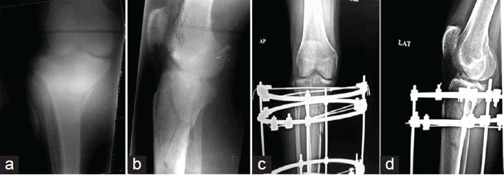
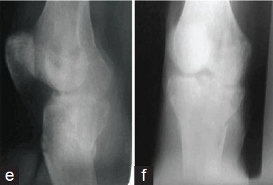
Functional outcome and gender were described using percentages and frequencies. An independent sample t-test was used to compare the two groups' VAS scores, length of stay in the hospital, and surgical time. The Chi-square test was used to compare the two groups' functional outcomes after six months. A p-value of 0.05 or less was considered to be significant.
This study included 104 patients with Schatzker's grade IV–VI fractures. Two groups of patients were created: Group A (using the ORIF procedure) and Group B (using the Ilizarov technique). There were 31 (59.6%) men and 21 (40.4%) women in group A, and 36 (69.2%) men and 16 (30.8%) women in group B. (Table 1)
Group A patients' mean age was 44.87±14.15 years, while group B patients' mean age was 44.25±15.68 years. 13(25.0%) of group A's members were in the 20–30 age range, whereas 12(23.1%) and 27(51.9%) belonged to the 31–45 and >45 age categories, respectively. Group B consisted of 11 (21.2%) people in the 20–30 age range, and 26 (50.0%) and 15 (288.8%) people in the 31–45 and >45 age ranges, respectively. (Table 2). The ORIF group's mean operative time was 182.02±36.53 minutes, while the Ilizarov group's mean was 160.25±21.57 minutes.
|
Table-1: Comparison of gender distribution between groups |
|||
|
Gender |
Groups |
Total |
|
|
ORIF technique |
Ilizarov technique |
||
|
Male |
31 |
36 |
67 |
|
59.6% |
69.2% |
64.4% |
|
|
Female |
21 |
16 |
37 |
|
40.4% |
30.8% |
35.6% |
|
|
Total |
52 |
52 |
104 |
|
100.0% |
100.0% |
100.0% |
|
This difference is statistically significant, with a p-value of 0.001 (Table-3). The average pain score in the ORIF group was 4.52±1.06 after 24 hours, compared to 3.40±1.21 in the Ilizarov group. This difference is statistically significant, with a p-value of 0.001. (Table-4) Hospital stays in the ORIF group averaged 6.85±1.88 days, while in the Ilizarov group they were 5.79±1.05 days. This difference is statistically significant, with a p-value of 0.001. (Table-5).
|
Table-2: Comparison of age distribution between groups |
|||
|
Age groups |
Groups |
Total |
|
|
ORIF technique |
Ilizarov technique |
||
|
20-30 years |
13 |
11 |
24 |
|
25.0% |
21.2% |
23.1% |
|
|
31-45 years |
12 |
15 |
27 |
|
23.1% |
28.8% |
26.0% |
|
|
>45 years |
27 |
26 |
53 |
|
51.9% |
50.0% |
51.0% |
|
|
Total |
52 |
52 |
104 |
|
100.0% |
100.0% |
100.0% |
|
|
Table-3: Comparison of operative time between groups |
|||||
|
Operative time (minutes) |
Groups |
n |
Mean |
Std. Deviation |
p-value |
|
ORIF technique |
52 |
182.02 |
36.53 |
0.001 |
|
|
Ilizarov technique |
52 |
160.25 |
21.56 |
||
|
Table-4: Comparison of pain score after 24hours post-operatively between groups |
|||||
|
VAS after 24 hours post-operatively |
Groups |
n |
Mean |
Std. Deviation |
p-value |
|
ORIF technique |
52 |
4.52 |
1.06 |
0.001 |
|
|
Ilizarov technique |
52 |
3.40 |
1.21 |
||
|
Table-5: Comparison of hospital stay between groups |
|||||
|
Hospital stay (days) |
Groups |
n |
Mean |
Std. Deviation |
p-value |
|
ORIF technique |
52 |
6.85 |
1.88 |
0.001 |
|
|
Ilizarov technique |
52 |
5.79 |
1.05 |
||
|
Table-6: Comparison of functional outcome between groups |
||||
|
Functional outcome |
Groups |
Total |
p-value |
|
|
ORIF technique |
Ilizarov technique |
|||
|
Excellent |
22 |
35 |
57 |
0.024 |
|
42.3% |
67.3% |
54.8% |
||
|
Good |
10 |
10 |
20 |
|
|
19.2% |
19.2% |
19.2% |
||
|
Fair |
16 |
5 |
21 |
|
|
30.8% |
9.6% |
20.2% |
||
|
Poor |
4 |
2 |
6 |
|
|
7.7% |
3.8% |
5.8% |
||
|
Total |
52 |
52 |
104 |
|
|
100.0% |
100.0% |
100.0% |
||
Functional outcome showed that, in the Ilizarov technique group, 35(67.3%) had excellent outcomes, while 10(19.2%), 5(9.6%), and 2(3.8%) had good, fair, and poor outcomes, respectively. In the ORIF technique group, 22(42.3%) had excellent outcomes, while 10(19.2%), 16(30.8%), and 4(7.7%) had good, fair, and poor outcomes, respectively. (Table-6).
The process of fracture displacement that happened at the time of injury is reversed in fracture reduction, which is the restoration of the proper anatomical location of fracture pieces. It comprises articular fragments in fractures with intra-articular involvement and the elevation of impacted cancellous bone. Reduction of extra-articular fractures involves restoring limb length, alignment, and rotation to allow the joints above and below the fracture to reposition themselves correctly. The process of reconstructing the typical three-dimensional spatial relationship between fracture pieces in relation to one another is known as fracture reduction. In clinical practise, this procedure can be carried out directly, indirectly, or frequently by combining the two methods.
The surgical technique, as described by Karl-Heinz Frosch et al., acknowledges the intricacy of tibial plateau fractures and the necessity of meticulous planning and execution. In order to get the best possible result for the patient, it emphasises the significance of exact fracture reduction and the adaptability to extend the surgical approach as needed. To guarantee a satisfactory outcome, this procedure probably calls for a high degree of surgical skill and expertise.14 According to research by Polat et al., plate osteosynthesis the use of plates to stabilise the fracture may produce better clinical outcomes than screw fixation in Schatzker types 2, 3, and 4 tibia plateau fractures. This suggests that some fractures may respond better to one approach than another. The text emphasises early knee motion and weight bearing following surgery, the use of grafts and internal fixation, the significance of surgical precision in treating tibial plateau fractures, and the selection of a fixation technique based on the particular type of fracture. 15 According to research by L. J. Gonzalez, knee pain usually stabilises a year after a tibial plateau fracture. Patients also continue to have gains in other areas of their functioning and health, as evidenced by their PROs and SMFA index scores. This emphasises how crucial continuous rehabilitation and long-term follow-up are to helping patients heal from tibial plateau fractures in the best way possible.16 Ahmad S. Naja et al. performed a meta-analysis on the topic of complete tibial plateau care. ExFix is more likely to cause OA and superficial infections. However, there are a number of contributing elements that need to be taken into account while treating complex tibial plateau fractures, including patient characteristics, surgical expertise, the cause of injury, and soft tissue integrity following the fracture. In addition to the mode of fixation, more research is required to evaluate the potential determining factors that may impact soft tissue integrity, postoperative reduction, and functional outcomes.17
Our study aimed to directly evaluate, to the best of our knowledge, the outcomes of patients treated in the same institution for proximal tibial fractures using ORIF versus external fixation. This has not been done before in the literature.
A study by Anil Murat Ozturk et al. found that the area of orthopaedics has benefited from the increased usage of 3D printing technology, particularly in the creation of patient-specific models based on real imaging data. The findings indicate that because 3D individual models are simple to construct, they can be used as treatment algorithm decision-makers. Using the 3D printing method, the appropriate preoperative planning, operational approach, and fixation may be chosen based on the morphological features of individual types of plateau fractures.18 According to a study by Gaunder CL et al., out of 102 patients, 16 of the participants experienced a surgical site infection, translating to a 15.7% infection rate. The study showed that an ASA classification of 3 or 4 is linked to a noticeably higher infection rate than an ASA classification of 1 or 2, highlighting the significance of managing patient co-morbidities during the peri-operative period. Higher ASA groups have an infection incidence of 23.7%, which is alarming and needs to be taken into consideration while making decisions. Surgeons should be aware of this problem, inform patients adequately about the relatively high risk, and investigate non-operative or minimally invasive therapy methods as alternatives to surgery.19 It is concluded from a study by Koushik Narayan Subramanyam et al. that in difficult tibial plateau fractures, an Ilizarov external fixator with or without minimum internal fixation delivers a satisfactory outcome. After the fixator is removed from Type VI Schatzker fractures, particular attention must be given to search for any slight loss of alignment. The Ilizarov technique is a good choice for treating difficult proximal tibial fractures when internal fixation and extensive dissection are not recommended because of soft tissue impairment and comminuted fractures. The Ilizarov approach allowed for the simultaneous treatment of bone loss, infection, non-union, and deformity.20 According to a study by Vivek Shetty et al., infections—both superficial and deep—are a common problem that have been documented in the literature to range from 2 to 28%. In cases with surgical proximal tibia fractures, literature reports a malunion rate ranging from 10 to 40 percent. According to some experts, the incidence of high velocity trauma ranges from 1% to 9%. The incidence of hardware irritation varies greatly and is estimated to be between 5 and 18%. In long-term series, 23–44 percent of cases are observed to have post-traumatic arthritis. Patients with proximal tibial fractures require subsequent surgery in about 9% of cases.21. Functional outcome analysis in this study showed that, among the groups using the ORIF technique, 22 (42.3%) had excellent outcomes, while 10 (19.2%), 16 (30.8%), and 4 (7.7%) had good, fair, and poor outcomes, respectively. In contrast, among the groups using the Ilizarov technique, 35 (67.3%) had excellent outcomes, while 10 (19.2%), 5 (9.6%), and 2 (3.8%) had good, fair, and poor outcomes, respectively. This analysis yielded a statistically significant p-value of 0.024. The stability of the knee is more important to the long-term prognosis than the amount of reduction achieved, while a less involved procedure may still produce acceptable outcomes. The preferred course of treatment is open reduction and internal fixation with the appropriate fixation devices if the fracture is displaced or depressed with joint instability. The randomised study design and sizeable sample size of 104 cases were the study's main advantages. In order to address the effect modifiers, a stringent exclusion criterion was adhered to, and the results were further stratified for age, gender, and other variables. One significant weakness of the current study was the absence of a comparison of skin and soft tissue issues between the two procedures, which is a crucial aspect that can reinforce the safety of this innovative approach. In the future, this kind of investigation should be prioritised in clinical research.
Compared to open reduction and internal fixation, Ilizarov external fixations are a more viable choice for fixing proximal tibial fractures, especially those that are difficult. Patients with complicated proximal tibial fractures respond better to the Ilizarov external fixation approach in terms of functional success than to the open reduction internal fixation technique. As long as the necessary equipment and personnel are available, it is therefore recommended that the Ilizarov external fixator be used in the care of such cases in future orthopaedic practise.
- Atif M, Mohib Y, Hasan OH, Rashid H. In the cost-conscious era: Ilizarov circular frame or uniplanar external fixator for management of complex open tibia shaft fracture, retrospective cohort study from a level-1 trauma center. JPMA. The Journal of the Pakistan Medical Association. 2020;70(2 (Suppl 1)):S20. https://doi.org/10.7759/cureus.5375
- Khare S, Patel P. To evaluate the fracture pattern and functional outcome of posterior tibial plateau fracture fixation. International Journal of Orthopaedics. 2019;5(2):700-5. https://doi.org/10.22271/ortho.2019.v5.i2j.78
- Dönmez G, Babayeva N, Torgutalp ŞŞ, Korkusuz F, Doral MN. Return to Play After Intra-articular Knee Fractures. Intraarticular Fractures: Minimally Invasive Surgery, Arthroscopy.2019:409-15. https://doi.org/10.1007/978-3-319-97602-0_44
- Shrestha R, Kandel M, Gupta HK, Shrestha SK, Dongol S, Hamal RR. A study of conservative management of tibial plateau fractures. Journal of College of Medical Sciences-Nepal. 2016 Mar 31;12(1):5-9. https://doi.org/10.3126/jcmsn.v12i1.14455
- Morasiewicz P, Burzyńska K, Orzechowski W, Dragan SŁ, Dragan SF, Filipiak J. Three-dimensional printing as a technology supporting the treatment of lower limb deformity and shortening with the Ilizarov method. Medical Engineering & Physics. 2018 Jul 1;57:6974 .https://doi.org/10.1016/j.medengphy.2018.04.009
- Gonzalez LJ, Hildebrandt K, Carlock K, Konda SR, Egol KA. Patient function continues to improve over the first five years following tibial plateau fracture managed by open reduction and internal fixation. The Bone & Joint Journal. 2020 May 1;102(5):632-7. https://doi.org/10.1302/0301-620X.102B5.BJJ-2019-1385.R1
- Gaunder CL, Zhao Z, Henderson C, McKinney BR, Stahel PF, Zelle BA. Wound complications after open reduction and internal fixation of tibial plateau fractures in the elderly: a multicentre study. International Orthopaedics. 2019 Feb 14;43:461-5. https://doi.org/10.1007/s00264-018-3940-9
- Shetty¹ V, Shekhar S, Wagh Y. Tibial Condyle Fractures: Current Concepts of Internal Fixation. Journal of Clinical Orthopaedics. 2021 Jan;6(1):32-44 https://doi.org/10.13107/jcorth.2021.v06i01.414.

An Official Publication of
Islamabad Medical & Dental College
Volume 12 Issue 4
Editorial
Address of Correspondence
Muhammad Farhan Farhat
Email:
mfarhan14@yahoo.com
Cite this article.Hassan K, Mahmood A, Khursheed A, Tasneem Z, Shafaat S, Khan F, Awais DK. Phenotypic Profile of Kidd Blood Group System in Northern Pakistani Population. J Islamabad Med Dental Coll. 2023; 12 (4): 276-281 DOI: https://doi.org/10.35787/jimdc.v12i4.1056