Pattern of Adenoviral Conjunctivitis Reported to Two Tertiary Care Hospitals During Epidemic in Islamabad, Pakistan
Afia Matloob Rana, Erum Yousafzai, Waseem Akhter, Saira Bano
Department of Ophthalmology, HBS Medical and Dental College, Islamabad
Objective: To determine clinical pattern of “adenoviral conjunctivitis” during its epidemic in Pakistan.
Methodology: A total of 370 patients diagnosed with “adenoviral conjunctivitis” were included in this descriptive study. Diagnosis was made by the combination of characteristic clinical features and a positive conjunctival swab PCR for “adenovirus”. Clinical pattern of “adenoviral conjunctivitis” was determined by documenting various patient characteristics. Data was analyzed using SPSS 22.
Result: Median age of the study participants was 28.50 (1.00 – 67.00) years. 195 (52.70%) were males and 175 (47.30%) were females. 190 (51.35%) patients had unilateral ocular involvement while 180 (48.65%) patients had bilateral ocular involvement. Median number of family members of patients that were infected simultaneously was 1 (0-5). Frequency of patients with “adenoviral conjunctivitis” who had concomitant corneal involvement was 30 (8.1%)
Conclusion: Epidemic of “adenoviral conjunctivitis” in Pakistan, fortunately, exhibit a milder and benign clinical pattern with higher prevalence in males and mostly involving single eye. In addition, only 8.1% patients have concomitant corneal involvement.
Keywords: Adenovirus, Conjunctivitis, Epidemic, Viral Conjunctivitis
Recently, an epidemic of ‘conjunctivitis’ has hit the population of Pakistan mainly affecting the big cities of the country leading to an increased load of patients on the ophthalmology outdoor departments. One of the major causes of “conjunctivitis” occurring in the form of epidemic is “adenovirus” which belongs to “Mastadenovirus” genus having approximately 51 different serotypes.1, 2Amongst all these serotypes, ‘adenovirus 8’, ‘adenovirus 19’ and “adenovirus 37” are the main culprits of causing infection in the form of an epidemic.3 In terms of prevalence, “adenovirus” infection constitutes for approximately 65% to 90% of the cases of ‘viral conjunctivitis’.4
Outbreaks of ‘adenovirus’ infections have been reported to occur on a global scale in recent times and it has been found that there are several factors that result in greater propensity of contracting this viral infection. The most important factors are over-crowding, lack of ventilation in the living environment and poor hygiene practices.5 There are several patterns by which ocular infection of “adenovirus can present clinically. “Epidemic keratoconjunctivitis (EKC)” is its most severe clinical form in which both conjunctiva and cornea become diseased leading to red eye, photophobia and pain in the diseased eye and have the potential to cause long-term damage and scarring to the affected eye. For this purpose, it is essential to timely diagnose this serious eye disease and provide early and appropriate management. Diagnosis of “adenoviral conjunctivitis” is made by obtaining a “conjunctival swab” from a patient having typical clinical features and performing PCR of the obtained swab to detect “adenovirus” using “Sanger’s method” of sequencing.6 Management of “adenovirus conjunctivitis” primarily involves supportive care but there are various agents that are used in practice with the aim to treat acute infection as well as management of its long-term morbidities. There is no FDA approved antiviral for treatment of adenoviral conjunctivitis. However, there are multiple options like topical steroids, topical NSAID’s, preservative free, artificial tears, povidone-iodine irrigation, topical cyclosporin and cold compresses. New treatments options include include sialic acid analogs, N-chlorotaurine and cold atmospheric plasma (CAP) 7,8 Whenever a country is hit by an epidemic, it is essential to carefully monitor its patterns in the local population as it proves to be very helpful in formulating a comprehensive plan not only to deal with the current manifestation of the epidemic but also for the future recurrences of the condition. Since, epidemic of “adenoviral conjunctivitis” is a major public health concern in the present time, it was essential to study its clinical pattern. Therefore, this study has been conducted with the aim of determining clinical pattern of ‘adenoviral conjunctivitis’ during its epidemic in Pakistan.
This descriptive cross-sectional study was conducted at the Ophthalmology Department of HBS medical and Dental College, and Rawal Institute of Health Sciences, Islamabad, during 13th August 2023 to 30th October 2023, after taking approval from the ethical review board .For calculation of appropriate sample size for the study, WHO sample size calculator was used by assuming “confidence interval of 95%”, “absolute precision of 3.85%” and ‘anticipated prevalence of adenoviral conjunctivitis of 17.7%’ 9 using following formula 10
Calculation gave the sample size of 370 patients which were selected for the study through “non-probability consecutive sampling technique’

Exclusion criteria: Patients with any other ocular surface disease patients, with bacterial conjunctivitis, patients who did not consent to be part of the study and those in which PCR of conjunctival swab was negative for “adenovirus” were excluded from the study.
Diagnosis of ‘adenoviral conjunctivitis’ was made by the combination of presence of characteristic clinical features (including redness of conjunctiva, photophobia, ocular pain, foreign body sensation and excessive tearing) and a positive conjunctival swab PCR for “adenovirus”, a method that have been used in various studies 11
After diagnosis and inclusion in the study, clinical demography of ‘adenoviral conjunctivitis’ was determined by documenting various patient characteristics including age (in years), gender, number of family
members infected, laterality of the disease and concomitant involvement of cornea. Corneal involvement was diagnosed by careful slit lamp examination to visualize “sub-epithelial infiltrates” and “punctate keratopathy”. All the patients were then given appropriate supportive therapy for their diseased eyes as per standard institutional protocol. “Data was analyzed by using Statistical Package for Social Sciences (SPSS) 22.00. Normality of data was checked by Shapiro-Wilk test which showed that age and number of family members infected were not normally distributed and were thus represented using median (IQR). Qualitative data (gender, laterality of the disease and involvement of cornea) was represented by using percentage and frequency.
A total of 370 patients diagnosed with “adenoviral conjunctivitis” were included in the study. Median age of the study participants was 28.50 (1.00 – 67.00) years. 195 (52.70%) of the patients were males and remaining 175 (47.30%) patients were depicted below in figure 1:
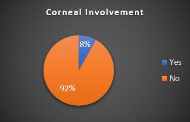
Figure 1: Frequency of concomitant corneal involvement in patients with “adenoviral conjunctivitis” (n = 370)
“Conjunctivitis” is a very common eye condition commonly encountered in ophthalmology clinics Stratification of clinical pattern by gender is tabulated in table I. Its pathophysiology is linked to a number of viruses, such as those belonging to the ‘Enterovirus’ genus and a variation of ‘Coxsackie virus A24’ but ‘adenoviruses’ are well recognized as
Table I: Stratification of clinical pattern by gender (n = 370) |
|||||
Parameter |
Male |
Female |
p-value |
||
Median age |
28 (1-67) years |
30 (2-62) years |
0.779 |
||
Median number of family members infected |
1 (0-4) |
1 (0-5) |
< 0.001 |
||
Laterality of the disease |
Unilateral |
110 (56.41%) |
Unilateral |
80 (45.71%) |
0.040 |
Bilateral |
85 (43.59%) |
Bilateral |
95 (54.29%) |
||
Concomitant involvement of cornea |
15 (7.69%) |
15 (8.57%) |
0.757 |
||
the primary etiological agents responsible for the onset of ‘acute conjunctivitis’.12,13
The primary manifestations of ocular ‘adenoviral conjunctivitis’ are due to abnormally valiant process of inflammation leading to redness of the eyes, excessive tearing, irritation, swelling, ‘epithelial keratitis’ and occasionally the presence of lymphadenopathy.14,15
The eye infection caused by adenovirus may exhibit rapid progression, with clinical signs being apparent approximately one week after the incubation period. Typically, the opposing eye may experience disease within a subsequent day, albeit with significantly diminished severity.16,17
In this study, it was observed that infection occurred predominantly in the male population. Similar findings were elaborated in a study conducted by Lee et al. 18 who reported that amongst all patients having ‘adenoviral conjunctivitis’ 58% were males. In addition, it was also observed that more than half of the patients of this study had unilateral ocular involvement which is congruent with the finding of a study in which it was reported in most of the cases of ‘adenoviral conjunctivitis’ were unilateral 19.
In this study, only 8.1% of the patients diagnosed with “adenoviral conjunctivitis” had concomitant corneal involvement. This frequency of corneal involvement was much lower as compared to a study conducted by Mohammed et al. 9 who reported that only 3.5% patients had normal cornea while having “adenoviral conjunctivitis” while all remaining patients had some degree of disease involving their cornea.
Similarly, in another study conducted by Pinto et al. 20 it was reported that frequency of corneal involvement in patients with ‘adenoviral conjunctivitis’ was 13.93% which was closer to what we found in our study. Contrarily, in another study conducted by Uemura et al. 21, it was reported that corneal involvement occurred only in 1.8% of the patients with “adenoviral conjunctivitis” which was much lower than what we observed in this study. Analysis of clinical pattern of the “adenoviral conjunctivitis” during epidemic in Pakistan shows that most patients have relatively benign condition with only a small proportion of patients having concomitant corneal involvement.
In addition, this study also illustrates the importance of conducting such surveys during an epidemic to ascertain and plan the future course of action in case of recurrence of the epidemic. However, limitation of our study include data collected from only two centers, we recommend multicenter study during epidemic to study the trend and pattern of disease.
Epidemic of ‘adenoviral conjunctivitis’ in Pakistan, fortunately, exhibit a milder and benign clinical pattern with higher prevalence in males and mostly involving single eye. In addition, only 8.1% patients have concomitant corneal involvement.
- Greber UF, Flatt JW. Adenovirus entry: from infection to immunity. Annu Rev Virol. 2019;6(1):177-197. https://doi.org/ 10.1146/annurev-virology-092818-015550.
- Wang J, Ning X, Xu Y, Wang R, Guo X, Xu J, et al. Etiological study of acute conjunctivitis caused by human adenovirus in Shanxi Province, China, between 2016 and 2019. Microbiol Spectr. 2023;11(4):e0015923. https://doi.org/10.1128/spectrum.00159-23.
- Imparato R, Rosa N, De Bernardo M. Antiviral drugs in adenovirus-induced keratoconjunctivitis. Microorganisms. 2022;10(10):2014. https://doi.org/10.3390/microorganisms10102014.
- Kuo IC, Gower EW. Cost savings from a policy to diagnose and prevent transmission of adenoviral conjunctivitis in employees of a large academic medical center. JAMA Ophthalmol. 2021;139(5):518-524 https://doi.org/10.1001/jamaophthalmol.2021.0150
- Islam M, Sultana ZZ, Iqbal A, Ali M, Hossain A. Effect of in-house crowding on childhood hospital admissions for acute respiratory infection: A matched case-control study in Bangladesh. Int J Infect Dis. 2021;105:639-645. https://doi.org/ 10.1016/j.ijid.2021.03.002.
- Thakur A, Goyal K, Chauhan P, Sharma B, Kant Dhir S, Katoch D, et al. Clinical presentation, investigation and control of an outbreak of adenoviral conjunctivitis in a neonatal unit at a tertiary hospital. Pediatr Infect Dis J. 2022;41(3):243-247. https://doi.org/ 10.1097/INF.0000000000003347.
- Labib BA, Minhas BK, Chigbu DI. Management of adenoviral keratoconjunctivitis: challenges and solutions. Clin Ophthalmol. 2020;14:837-852. https://doi.org/10.2147/OPTH.S207976.
- Aljohani RM, Mohammed FAG, Abu Tuwaymah AMA, Althubiany MSA, Abdulghani Khalil AM. Epidemic keratoconjunctivitis; a general review of the disease, its management and prevention recommendations. Int J Med Dev Ctries. 2021;5(2):747-750. https://doi.org/10.24911/IJMDC.51-1608123469.
- Mohammed MA, Khalil MA, Fayyadh RA. Prevalence and clinical features of ocular adenoviral infection among patients attended to Ibn-Alhaitham teaching eye hospital in Baghdad, Iraq: A molecular study. Med Legal Update. 2020;20(4):931-937. https://doi.org/10.37506/mlu.v20i4.1942.
- Naing L, Nordin RB, Abdul Rahman H, Naing YT. Sample size calculation for prevalence studies using Scalex and ScalaR calculators. BMC Med Res Methodol. 2022;22(1):209 https://doi.org/10.1186/s12874-022-01694-7.
- Martin C, Löw U, Quintin A, Schießl G, Gärtner B, Heim A, Seitz B. Epidemic keratoconjunctivitis: efficacy of outbreak management. Graefes Arch Clin Exp Ophthalmol. 2022;260(1):173-180. https://doi.org/10.1007/s00417-021-05344-4.
- Shorter ES, Whiteside MM, Harthan JS, Morettin CE, Perera CD, Johnson SD, et al. Diagnostic accuracy of clinical signs, symptoms and point-of-care testing for early adenoviral conjunctivitis. Clin Exp Optom. 2022;105(7):702-707. https://doi.org/10.1080/08164622.2021.1984180.
- Muto T, Imaizumi S, Kamoi K. Viral conjunctivitis. Viruses. 2023;15(3):676. https://doi.org/10.3390/v15030676.
- Kuo IC. Adenoviral keratoconjunctivitis: diagnosis, management, and prevention. Curr Ophthalmol Rep. 2019; 7:118–127. https://doi.org/: 10.1007/s40135-019-00207-y.
- Hoffman J. Adenovirus: ocular manifestations. Community Eye Health. 2020;33(108):73-75.
- Koçluk Y, Alyamaç Sukgen E. Different clinical presentations of the same epidemic keratoconjunctivitis outbreak in premature babies and their parents. Ocul Immunol Inflamm. 2018;26(3):406-411. https://doi.org/10.1080/09273948.2016.1219751.
- Rajaiya J, Saha A, Ismail AM, Zhou X, Su T, Chodosh J. Adenovirus and the cornea: more than meets the eye. Viruses. 2021;13(2):293. https://doi.org/10.3390/v13020293.
- Lee CS, Lee AY, Akileswaran L, Stroman D, Najafi-Tagol K, Kleiboeker S, Chodosh J, Magaret A, Wald A, Van Gelder RN; BAYnovation Study Group. Determinants of outcomes of adenoviral keratoconjunctivitis. Ophthalmology. 2018;125(9):1344-1353. https://doi.org/10.1016/j.ophtha.2018.02.016.
- Meyer-Rüsenberg B, Loderstädt U, Richard G, Kaulfers PM, Gesser C. Epidemic keratoconjunctivitis: the current situation and recommendations for prevention and treatment. Dtsch Arztebl Int. 2011;108(27):475-480. https://doi.org/10.3238/arztebl.2011.0475.
- Pinto RD, Lira RP, Arieta CE, Castro RS, Bonon SH. The prevalence of adenoviral conjunctivitis at the Clinical Hospital of the State University of Campinas, Brazil. Clinics (Sao Paulo). 2015;70(11):748-750 https://doi.org/10.6061/clinics/2015(11)06.
- Uemura T, Migita H, Ueno T, Tsukahara-Kawamura T, Saeki Y, Fujimoto T, Uchio E. Clinical and virological analysis of epidemic keratoconjunctivitis caused by adenovirus type 54 in a regional ophthalmic clinic in Kyushu, Japan. Clin Ophthalmol. 2018; 12:511-517 https://doi.org/10.2147/OPTH.S148264.

An Official Publication of
Islamabad Medical & Dental College
Volume 13 Issue 1
Address of Correspondence
Afia Matloob Rana
Email:
afiamatloob@yahoo.com
Authors’ Contribution: 1,2Conception; Literature research; manuscript design and drafting; 2,3 Critical analysis and manuscript review; 4Data analysis; Manuscript Editing.
Cite this article. Rana AM, Yousafzai E, Akhter W, Bano S. Pattern of adenoviral conjunctivitis reported to two tertiary care hospitals during epidemic in Islamabad, Pakistan. J Islamabad Med Dental Coll. 2024; 13(1): 34-38.DOI: https://doi.org/10.35787/jimdc.v13i1.1066