Prevalence of Interstitial Lung Disease in Patients with Chronic Cough Taking Chest X-rays and CT Chest as Diagnostic Tools
Abdul Qadeer Khan 1, Shaista Nayyar1, Sohaib Khalid1, Irum Taqi1, Murtaza Ahmed Khan2
1Department of Radiology, PAF Hospital, Islamabad; 2Department of Neurosurgery, Sir Ganga Ram Hospital, Lahore
Objective: To assess the prevalence of ILD among chronic cough patients and to compare the diagnostic accuracy of CT and CXR.
Methodology: A cross-sectional study on 200 patients was performed in Pakistan Air Force Hospital, Islamabad for a period of 6 months from 15th April to 15th October, 2023. Chest X-ray (CXR) and Computed Tomography (CT) Chest were used as diagnostic tools.
Results: The chest x-ray detected ILD in 114 (57%) while the chest CT identified in 178 (89%) of the patients. CXR and CT had a sensitivity of 65.05 and 95.65 and a specificity of 45.1 and 85.2 respectively. CXR and CT detected reticular opacity, nodular opacity, and ground glass opacity in 66% vs 81%, 41% vs 78%, and 33% vs 55% of the ILD patients. Dyspnoea (85%), fever (30%), arthralgia (24%), and weight loss (9%) were found as clinical indications.
Conclusion: CXR can be used to perform initial preliminary tests to filter out the presence of other diseases in patients of ILD. However, it cannot be solely relied on as a diagnostic tool. The study aimed at assessing the prevalence of ILD among chronic cough patients while comparing the diagnostic accuracy of CT and CXR. CT Chest can be the choice of imaging modality in diagnosing ILD as it outperformed CXR in many aspects of our study, with better precision, accuracy, sensitivity, and specificity.
Keywords: Interstitial Lung Disease, Chronic Cough, Chest X-ray, Computed Tomography
Cardiovascular Interstitial lung disease (ILD) is a broad term that encompasses multiple disorders ranging from occasional inflammatory processes to severe non treatable lung fibrosis. Classically ILD produce the three Cs’: cough, clubbing of nails and coarse crackles on auscultation.1 The similarity in imaging reports along with the combination of diseased indications classify them into further subdivisions2. They are predominantly characterized by inflammation or fibrosis of the lung interstitium3. This primarily affects the normal functioning of the lungs and hampers the person’s breathing, eventually deteriorating the overall quality of life4. A multitude of factors, principally the worsening air quality with the passage of time has caused a global rise in the incidence and prevalence of ILD, with about 32 and 71 persons per 100,000 respectively5,6. The unfavorable interaction of some pharmaceuticals and emissions of harmful gases, smoking as well as organic/inorganic dusts is also reported to add to the progression of ILD.
Cough, dyspnea, phlegm, post-nasal drip, weight loss, and dysphagia are some of the common symptoms of ILD with particular reference to the Pakistani population7. The typical imaging features on HRCT chest include the presence of reticular opacities, centrilobular nodules, interlobular septal thickenings and soft tissue haziness which occurs variably in different forms of ILDs. The most frequently presenting symptom however is chronic cough with an occurrence in around 80% of ILD patients8. Even globally, it is generally frequent in 1 out of 10 adult population9,10. Although cough is a natural reflex mediated with the beneficence of aspiration prevention, it badly affects the quality of life11. Chronic cough lasts around sixty days with increased chest pain and frequent sleep disturbances12,13. It is usually assessed through Leicester Cough Questionnaire (LCQ), a subjective quantifiable method14.
The clinical signs of ILD are often mistakenly taken for other ordinary diseases of the lung. Therefore, diagnosis of ILD is quite challenging and the accuracy rates with conventional tests are quite low 15. Even after a detailed examination including surgical lung biopsy (SLB), around 12% of the patients remain undiagnosed16. Only the correct diagnosis can lead to a better management plan that is tailored to individual patient needs and can result in the quick recovery of the patient from an ailing state back to a normal healthy life17.
Therefore, with the growing health burden caused by ILD, the role of early diagnosis through regular imaging of the chest with quality diagnosis is crucial 18. Imaging tools like chest X-rays and CT chests augments the informed decision-making of clinicians regarding better treatment options19. However, there is a lack of empirical data on the prevalence of interstitial lung disease in patients with chronic cough through using chest X-rays (CXR) and CT chest as diagnostic tools. Therefore, this study is being performed to assess and understand this phenomenon.
A cross-sectional study was performed in Pakistan Air Force Hospital, Islamabad for a period of 6 months from 15th April to 15th October. The study was approved by the institutional ethical review board and detailed informed consent from the study participants was taken. The patients and their attendants were then informed in detail about the objectives of this study along with the procedures involved.
This research involved a total of 200 patients referred to the Department of Radiology from the pulmonary medicine OPD. Keeping into account the patient’s socio-economic history and after physical examinations, and pulmonary function tests (PFTs), they were assessed for the presence of Interstitial Lung Disease by using a chest X-ray followed by computed tomography (CT) chest to substantiate the previous findings. The GE and Med equips X-ray machines with tube current of 500 and 300 milliamperes respectively; whereas, Toshiba 160 slice CT scan machine with tube voltage of 120 kV and tube current modulation between 120-250mA was being used in our study. The HRCT protocol included an un enhanced image of the thorax (slice thickness 2mm and slice interval 1mm) acquired with a standard kernel and soft tissue window and reconstruction of the parenchyma with a sharp kernel and lung window.
The data related to patient profiles was recorded and organized on a questionnaire while maintaining confidentiality and security. The PA view x-ray of the chest was performed at 55 kVp. Study participant’s characteristics that could be quantified like weight or age were computed through mean ± standard deviation. Whereas study participants’ characteristics that could be categorized like the presence or absence of ILD were determined through frequencies or percentages. The sensitivity and specificity of the HRCT and X-ray was determined by the following Sensitivity= TP/ (TP+FN) and Specificity= TN/(TN+FP). Abbreviations: TP, true positive; TN, true negative; FP, false positive; FN, false negative. Correlations were made using chi-square tests. The imaging reports and other findings were assessed and analyzed on Excel and SPSS version 26.
Inclusion criteria: ILD-suspected patients from both the male and female genders were included. These were referred to the Radiology department with chronic cough and afterward were found to have the following chest imaging results: Ground-glass opacity (GGO), honeycombing (presence of cysts and collagen fibers; last-stage ILD), bilateral reticular or reticulonodular opacities, clear and indubitable chest radiographs, pleural-parenchymal edges along the hemi diaphragm and heart weakly distinguished, dilatation of bronchioles.
Exclusion criteria: Patients with ischemic heart disease, tuberculosis, chronic liver/kidney diseases, pregnant women and any patient < 18 years were excluded from the study.
This The 200 participants of our study had a mean age of 42.7±.11.3. The subjects in our study had an age ranging from 20 years to 73 years. 142 (71%) patients were males and 58 (29%) were females whose symptoms lasted 8.3±1.9 years (figure 1).
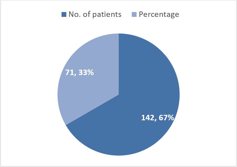
Figure 1 Gender distribution of subjects
The chest x-ray detected ILD in 114 (57%) while the chest CT identified it in 178 (89%) of the patients
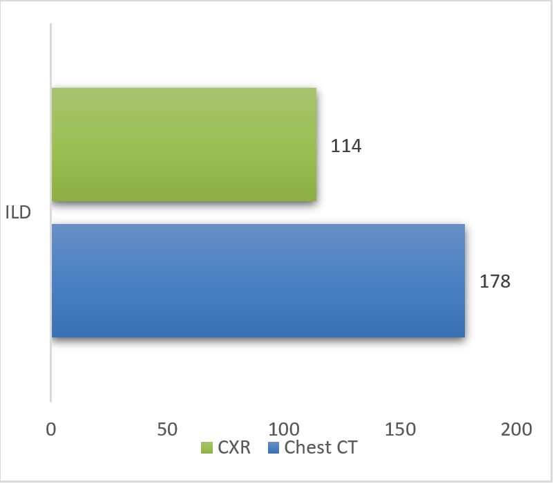
Figure 2 Comparison of CXR with CT in ILD detection
(figure 2). The sensitivity of CXR was 65.05 whereas the CT had a sensitivity of 95.65. The specificity of CXR was 45.1 whereas the CT had a specificity of 85.2 (Table 1).
Table I: Diagnostic value of CXR vs CT |
|||
|
Diagnostic value |
||
|
|
Sensitivity |
Specificity |
CXR |
65.05 |
45.1 |
|
CT Chest |
95.65 |
85.2 |
|
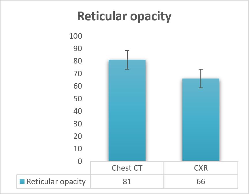
Figure 3 Detection of reticular opacity in CXR vs CT
Nodular opacities were detected in 41% of patients through CXR whereas the CT had a rate of 78%, statistically significant, P<0.05 (figure 4).
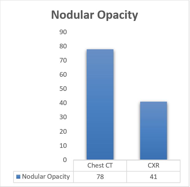
Figure 4 Detection of nodular opacity in CXR vs CT
Honeycombing was detected in 21% of patients through CXR whereas CT detected it in 43% of patients (figure 5).
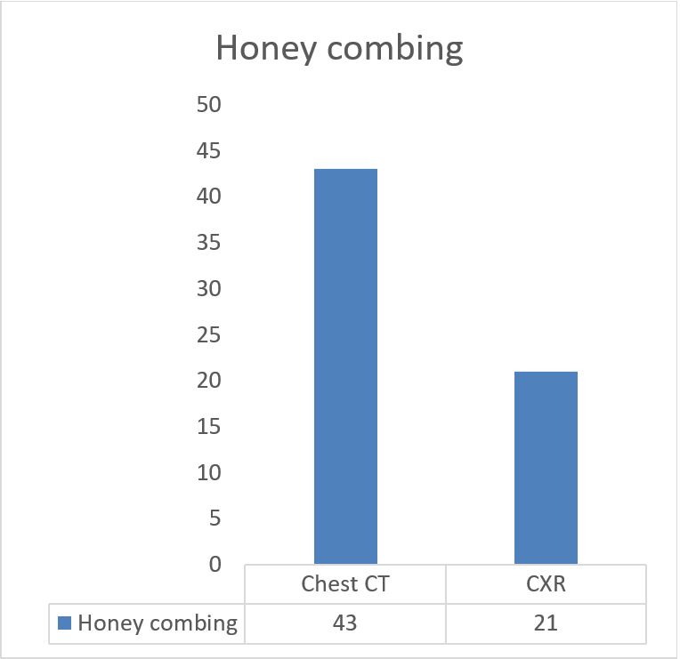
Figure 5 Detection of honey combing in CXR vs CT
Ground Glass Opacity was detected in 33% of patients through CXR whereas the CT detected in 55% patients (Figure 6).
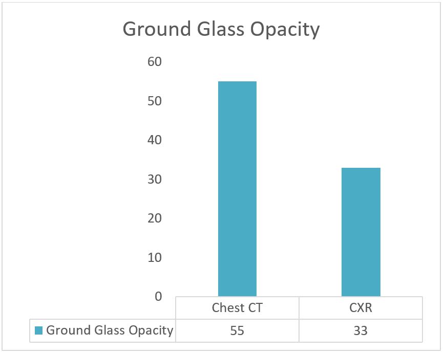
Figure 6 Ground glass opacity in CXR vs CT
We conducted this study to assess the prevalence of interstitial lung disease in patients with chronic cough taking chest x-rays and CT chest as diagnostic tools. Different symptoms and clinical anomalies were used to evaluate and compare these imaging tools.
ILD primarily targets males which was evident in our study as well where 71% of the patients were males 20. In our study subjects, the symptoms lasted 8.3±1.9 years. Similarly, a study in Ayub Teaching Hospital reported their figures to be 9.47±1.7 years21. HRCT is the gold standard for the diagnosis of ILDs whereas we compared the accuracies of both the CT and CXR. The study found that the overall performance of CT was better than CXR in terms of diagnosing ILD. The chest x-ray detected ILD in 114 (57%) which was quite similar to the 58.39% of the
patients diagnosed in a study conducted by Afzal et al in the Combined Military Hospital (CMH), Rawalpindi. On the contrary, the chest CT identified it in 178 (89%) of the patients. The ability of the imaging test to diagnose a patient (positive) was found to be superior in CT. The sensitivity of CXR was 65.05 whereas the CT had a sensitivity of 95.65. Similarly, the ability of the imaging test to diagnose a healthy individual (negative) was found to be superior in CT. The specificity of CXR was 45.1 whereas the CT had a sensitivity of 85.2. These results are in line with the findings of other studies. Walsh et al in the European Respiratory Review ruled in favor of computed tomography tests over X-rays due to their higher sensitivity and specificity 22.
A descriptive study conducted by Agrawal et al found that the presence of ILD went undiagnosed through the conventional chest radiographs. 23 Although we did not perform surgical lung biopsy in our study, the literature review suggests that CXR fails to detect around 10-12 % of those ILD patients in whom ILD is detected after SLB16,24. In terms of spotting pulmonary variabilities, the CT chest had a better response. Reticular opacity was most frequently detected in our patients as 66% of patients through CXR and 81% of patients through CT were detected. Two cross-sectional studies by Remi-Jardin et al and Patel et al also found a related reticular pattern to be the most frequent observation, with 37% / 63% and 50% / 70% results in CXR / CT respectively.25,26 All three of these studies point towards higher sensitivity of CT over CXR. Nodular opacities were the second most common finding in 41% of patients through CXR whereas in 78% through CT, statistically significant (P<0.05). Ground Glass Opacity (GGO) was detected in 33% of patients through CXR whereas CT detected in 55% of patients.
The prevalence of ILD is far more common in patients with the history of chronic cough along with other typical symptoms with male predominance. Although the chest x-ray is a simple test, costs low, and is easily accessible even in poorly equipped healthcare setups, it is not the gold standard in diagnosing high-end diseases like ILD. CXR can, however, be used to perform initial preliminary tests to filter out the presence of other diseases. The ability of the imaging test to diagnose a patient (positive) was found to be superior in CT. Similarly, the ability of the imaging test to diagnose a healthy individual (negative) was found to be superior in CT. CT Chest is therefore the choice of imaging modality to diagnose ILD and other pulmonary anomalies with better precision, accuracy, sensitivity, and specificity.
- Wijsenbeek M, Suzuki A, Maher TM. Interstitial lung diseases. The Lancet. 2022;400(10354):769-786. https://doi.org/10.1016/S0140-6736(22)01052-2
- Kaul B, Cottin V, Collard HR, Valenzuela C. Variability in Global Prevalence of Interstitial Lung Disease. Front Med (Lausanne). 2021;8. https://doi.org/10.3389/fmed.2021.751181
- Troy LK, Grainge C, Corte TJ, et al. Diagnostic accuracy of transbronchial lung cryobiopsy for interstitial lung disease diagnosis (COLDICE): a prospective, comparative study. Lancet Respir Med. 2020;8(2):171-181. https://doi.org/10.1016/S2213-2600(19)30342-X
- Wong AW, Ryerson CJ, Guler SA. Progression of fibrosing interstitial lung disease. Respir Res. 2020;21(1):32. https://doi.org/10.1186/s12931-020-1296-3
- Kaul B, Cottin V, Collard HR, Valenzuela C. Variability in Global Prevalence of Interstitial Lung Disease. Front Med (Lausanne). 2021;8. https://doi.org/10.3389/fmed.2021.751181
- Shah AK, Kushwah APS, Pandey S, Tomar SP. Role of HRCT in Interstitial Lung Disease with Radiographic Correlation. Journal of Evidence Based Medicine and Healthcare. 2020;7(44):2573-2578. https://doi.org/10.18410/jebmh/2020/531
- Jafri S, Ahmed N, Saifullah N, Musheer M. Epidemiology and Clinico-radiological features of Interstitial Lung Diseases. Pak J Med Sci. 2020;36(3):365-370. https://doi.org/10.12669/pjms.36.3.1046
- Van Manen MJG, Hart SP. The Curious Case of Cough in Interstitial Lung Diseases. Chest. 2022;162(3):501-502.
- Song WJ, Morice AH. Cough Hypersensitivity Syndrome: A Few More Steps Forward. Allergy Asthma Immunol Res. 2017;9(5):394. https://doi.org/10.4168/aair.2017.9.5.394
- Song WJ, Chang YS, Faruqi S, et al. The global epidemiology of chronic cough in adults: a systematic review and meta-analysis. European Respiratory Journal. 2015;45(5):1479-1481.
- .Lee KK, Matos S, Evans DH, White P, Pavord ID, Birring SS. A Longitudinal Assessment of Acute Cough. Am J Respir Crit Care Med. 2013;187(9):991-997. https://doi.org/10.1164/rccm.201209-1686OC
- Lan NSH, Moore I, Lake F. Understanding cough in interstitial lung disease: a cross‐sectional study on the adequacy of treatment. Intern Med J. 2021;51(6):923-929. https://doi.org/10.1111/imj.14837
- Van Manen MJG, Wijsenbeek MS. Cough, an unresolved problem in interstitial lung diseases. Curr Opin Support Palliat Care. 2019;13(3):143-151. https://doi.org/10.1097/SPC.0000000000000447
- Birring SS, Kavanagh JE, Irwin RS, et al. Treatment of Interstitial Lung Disease Associated Cough. Chest. 2018;154(4):904-917.
- Walsh SLF, Wells AU, Desai SR, et al. Multicentre evaluation of multidisciplinary team meeting agreement on diagnosis in diffuse parenchymal lung disease: a case-cohort study. Lancet Respir Med. 2016;4(7):557-565.
- Guler SA, Ellison K, Algamdi M, Collard HR, Ryerson CJ. Heterogeneity in Unclassifiable Interstitial Lung Disease. A Systematic Review and Meta-Analysis. Ann Am Thorac Soc. 2018;15(7):854-863.
- Meyer KC. Diagnosis and management of interstitial lung disease. Transl Respir Med. 2014;2(1):4.
- George PM, Spagnolo P, Kreuter M, et al. Progressive fibrosing interstitial lung disease: clinical uncertainties, consensus recommendations, and research priorities. Lancet Respir Med. 2020;8(9):925-934.
- Meyer KC, Raghu G. Patient Evaluation. In: Diffuse Lung Disease. Springer New York; 2012:3-16. https://doi.org/10.1007/978-1-4419-9771-5_1
- Senger KPS, Singh A. High-resolution computed tomography as a non-invasive imaging biomarker for interstitial lung diseases: a tertiary center study. International Journal of Advances in Medicine. 2021;8(2):207. https://doi.org/10.18203/2349-3933.ijam20210265
- Akram F, Hussain S, Azmat A, Javed H, Fayyaz M, Ahmed K. Diagnostic Accuracy Of Chest Radiograph In Interstitial Lung Disease As Confirmed By High Resolution Computed Tomography (HRCT) Chest. Journal of Ayub Medical College Abbottabad. 2022;34(4(SUPPL 1)):1008-1012. https://doi.org/10.55519/JAMC-04-S4-11183
- Walsh SLF, Devaraj A, Enghelmayer JI, et al. Role of imaging in progressive-fibrosing interstitial lung diseases. European Respiratory Review. 2018;27(150):180073. https://doi.org/10.1183/16000617.0073-2018
- Agrawal MK, Kumar A, Agrawal R, Rana R. To study the significance of HRCT over Chest X-ray in the diagnosis of interstitial lung diseases. J Evol Med Dent Sci. 2019;8(2):94-98. https://doi.org/10.14260/jemds/2019/21
- Hodler J, Kubik-Huch RA, von Schulthess GK, eds. Diseases of the Chest, Breast, Heart and Vessels 2019-2022. Springer International Publishing; 2019. https://doi.org/10.1007/978-3-030-11149-6
- Patel T, Ilangovan G, Balganesan H. Comparative Study of X-ray and High Resolution Computed Tomography in the Diagnosis of Interstitial Lung Disease. International Journal of Contemporary Medicine, Surgery and Radiology. 2020;5(1).
- Remy-Jardin M, Remy J, Deffontaines C, Duhamel A. Assessment of diffuse infiltrative lung disease: comparison of conventional CT and high-resolution CT. Radiology. 1991;181(1):157-162.

An Official Publication of
Islamabad Medical & Dental College
Volume 13 Issue 1
Address of Correspondence
Abdul Qadeer Khan
Email:
abdulqadeerkhan8331@gmail.com
Authors’ Contribution: 1,2Conception; Literature research; manuscript design and drafting; 3,4 Critical analysis and manuscript review; 5Data analysis; Manuscript Editing.
Cite this article. Khan AQ, Nayyar S, Khalid S, Taqi I, Khan MA. Prevalence of Interstitial Lung Disease in Patients with Chronic Cough Taking Chest X-rays and CT Chest as Diagnostic Tools. J Islamabad Med Dental Coll. 2024; 13(1): 110-115.
DOI: https://doi.org/10.35787/jimdc.v13i1.1073.