Systemic Lupus Erythematosus (SLE) Presenting as Mixed Sensori-Motor Axonal Polyneuropathy
Mutahra Khaliq1, Nauman Ismat Butt2, Muhammad Sohail Ajmal Ghoauri1, Fahad Qaisar1, Tayyab Hashmi1, Sabir Ali1
1Bahawal Victoria Hospital, Quaid-e-Azam Medical College, Bahawalpur, Pakistan, 2Azra Naheed Medical College, Superior University Lahore Pakistan
Systemic Lupus Erythematosus (SLE) is an autoimmune disease that has multisystem manifestations. However, axonal polyneuropathy is rare in SLE. We report the case of a 41-years old lady who presented with gradual onset, progressively worsening bilateral lower limb weakness for one month along with tingling, prickling and burning sensation. There was history of dry eyes, oral ulcers, dry mouth, dental caries, lethargy and fatigue. On examination, poor oral hygiene, coarse dry tongue, oral ulcers, dental caries and conjunctival pallor were noted. Neurological examination of lower limbs revealed flaccid paralysis with paresthesia of both feet extending up to the mid-shin level. She had normochromic normocytic anemia with hemoglobin 10.8 g/dl and raised ESR 40 mm/hour. NCS revealed mixed sensori-motor axonal polyneuropathy. ANA, anti-DsDNA (138 IU/ml), Anti-Sm (>200 U/ml) and anti-Ro/SSA antibodies (146 U/ml) were positive with low serum C3 (29.3 mg/dl) and low serum C4 (07 mg/dl). She was diagnosed as SLE and started on plasmapheresis, steroids, hydroxycholoroquine and azathioprine with marked improvement at follow-up.
Keywords: Axonal, Azathioprine, Hydroxycholoroquine, Plasmapheresis, Prednisolone, Systemic Lupus Erythematosus (SLE).
Systemic Lupus Erythematosus (SLE) is an autoimmune disease that has multi-system manifestations that may vary from mild muco-cutaneous disease to severe and extensive multi-organ involvement resulting in musculo-skeletal, renal, cardiac and neuropsychiatric disease. Timely detection and advances in management has significantly reduced SLE related mortality and morbidity has reduced significantly. The 10-year survival rate of SLE was 95% in 2000s which shows a significant reduction from 63.2% rate in 1950s.1 However delay in diagnosis and inadequate disease control leads to significant disease burden and mortality. The prevalence of neuropsychiatric involvement in SLE varies from 37% to 90%.2 Neuropsychiatric involvement in SLE is usually because of central nervous system disease such as cerebritis3 but rarely may be caused by peripheral nervous system. Unterman et al.4 reported neuropsychiatric manifestations to be present in 56.3% patients with SLE but polyneuropathy and mononeuropathy were seen in only 1.5% and 0.9% patients of SLE respectively. Xianbian et al.2 reported peripheral neuropathy in 1.5% of 4924 SLE patients included, with polyneuropathy being the most common neuropathy seen in 0.9%.2 Furthermore, the association of SLE with axonal variants of neuropathy has been reported rarely. Florica et al.5 demonstrated that patients of SLE with peripheral neuropathy had significantly decreased age and sex standardized physical component summary scores (SF-36 questionnaire and health-related quality of life) leading to a lower quality of life as compared to patients of SLE without neuropathy. Even though neuropathy may not be linked to increased mortality directly. Moreover, there are no standard clinical guidelines available regarding management of this condition. Treatment is usually tailored according to severity of neuropathy and other systems involved on a case-to-case basis.6
Case Report:
A 41-years old lady presented with bilateral lower limb weakness for 1 month. It was of insidious onset, gradually worsening, initially causing slight disturbance in walking but progressed to the extent that the patient could not mobilize and became bed bound for last 5 days. Along with these symptoms, there was history of tingling, prickling and burning sensation of both feet extending up till the shins. There was no history of trauma, fever, urinary or fecal incontinence, fits, altered sensorium, visual and auditory disturbances or previous episodes of limb weakness. However, on exploration of history, there was history of grittiness and dryness of eyes, recurrent oral ulcers, dry mouth, repeated dental caries, generalized lethargy and fatigue. Joint pain without redness or swelling involving the small joints of hands and wrists were reported associated with morning stiffness lasting 30-60 minutes. No history of genital ulcers, photosensitivity, skin rashes, alopecia, renal stones, puffy hands or Raynaud’s phenomena was reported. She did not smoke or use illicit drugs. She was married with 4 children and had no history of miscarriages. There was no family history of neurological or rheumatic diseases. The patient had consulted a few local GPs with no relief of symptoms. On examination, the patient was conscious and cooperative with poor oral hygiene, coarse dry tongue, 2 oral ulcers on the lower buccal surface, multiple dental caries and conjunctival pallor. Tenderness without swelling was present at both wrists, both ankles and multiple proximal interphalangeal and metacarpo-phalangeal joints bilaterally. There was no cyanosis, clubbing, pedal edema, lymphadenopathy or parotid swelling. Neurological examination of lower limbs revealed normal muscle tone, reduced power (grade 2/5), absent knee and ankle reflexes with downgoing plantar response bilaterally. There was paresthesia of both feet extending up to the mid-shin level. Upper limbs demonstrated normal sensory and motor examinations. There was no cerebellar or cranial nerve involvement. Examination of precordium, chest and abdomen were unremarkable. She was admitted for work-up of flaccid paralysis. On investigation, CBC revealed normochromic normocytic anemia with hemoglobin 10.8 g/dl and normal TLC and platelet counts. ESR was raised at 40 mm/hour with normal CRP. RFTs, LFTs, serum electrolytes including potassium, serum TSH and urinalysis were normal. Serologies for Syphilis, HIV, Hepatitis B and C were negative. Serum aldolase and CPK were normal. As shown in Figure 2 and 2, Nerve conduction studies (NCS) revealed mixed sensori-motor axonal polyneuropathy in the lower limbs bilaterally with normal upper limbs. Trans-thoracic echocardiography and ultrasound abdomen & pelvis were within normal parameters. The Schirmer test revealed 6 mm and 8 mm wetting of filter paper strip after 5 minutes for right eye left eye respectively. An autoimmune profile was ordered which revealed positive ANA, anti-DsDNA (138 IU/ml), Anti-Sm (>200 U/ml) and anti-Ro/SSA antibodies (146 U/ml) with low serum C3 (29.3 mg/dl) and low serum C4 (07 mg/dl) but negative RA factor, c-ANCA, p-ANCA, anti-Jo-1 and anti-RNP antibodies. Based on clinical and laboratory findings, a diagnosis of mixed axonal polyneuropathy due to Systemic Lupus Erythematosus (SLE) was made. She was started on pulse intravenous methylprednisolone 1000mg/day for 5 days followed by oral prednisolone (1mg/kg body weight per day) with gradual dose tapering.
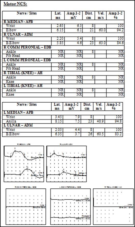
Figure 1: Motor Nerve Conduction Studies showing no response in bilateral common peroneal and tibial nerves with normal response in nerves of upper limbs
Five sessions of plasmapheresis were done on alternate days which resulted in resolution of paresthesia, improvement of power to grade 4/5 and the patient was able to walk with assistance. In addition, she was prescribed hydroxycholoroquine (5mg/kg body weight per day), azathioprine (1mg/kg body weight per day), sunblock SPF 60, calcium and vitamin D supplements, oral essomeprazole and artificial tear eye drops. On follow up at 12 weeks, the patient had fully improved with power 5/5 in both lower limbs. She was tolerating hydroxycholoroquine (5mg/kg/day), azathioprine (2mg/kg/day) and prednisolone (5mg/day) without any adverse effects.
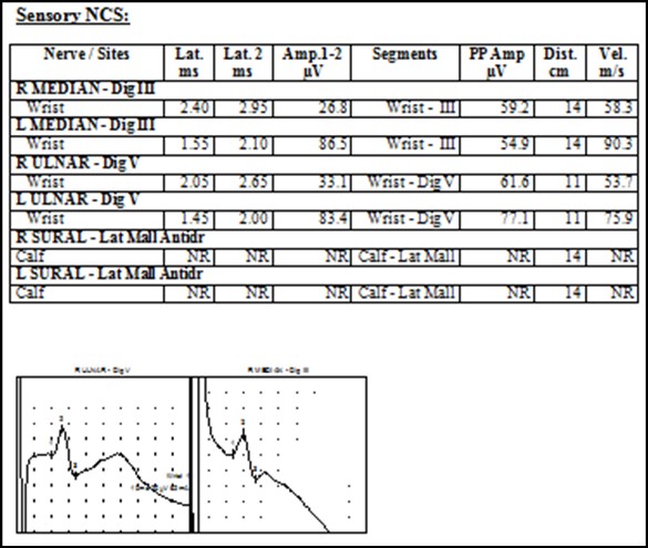
Figure 2: Sensory Nerve Conduction Studies showing no response in bilateral sural nerve with normal response in nerves of upper limbs
Even though the outcome of SLE patients has improved drastically over the years, SLE may still lead to substantial disease burden and increased death risk. The common causes of mortality in SLE patients include infections (24.5%), atherosclerosis (15.7%), disease flare (13.3%) and malignancies (9.6%).7 Lupus patients with disease onset before the age of 40 years were three times more likely to die of any cause compared to those without lupus.7 Therefore timely diagnosis and adequate treatment of SLE is vital. Having a specificity and sensitivity of 92% and 97% respectively, the Systemic Lupus International Collaborating Clinics (SLICC) criteria is usually employed to diagnose SLE.8 SLE is diagnosed in presence of >4 criteria, at least 1 immunological criteria and 1 clinical criteria.8 On basis of the SLICC criteria, our patient had a score of 7: oral ulcers, arthralgias, peripheral neuropathy, positive ANA, positive Anti-DsDNA & positive Anti-Sm antibodies and low serum complement (C3 and C4). Pathogenesis of peripheral neuropathy in SLE patients is not fully understood. Possible mechanisms include small nerve fiber injury, immune complex deposition, small vessel disease, Vasculitis and antibodies-associated inflammation.2 Mahler et al.9 reported anti-Sm antibodies to be associated with higher risk of peripneral neuropathy. Harel et al.10 confirmed presence of anti-cardiolipin antibodies in SLE patients with neuropathy. Our patient with neuropathy had positive anti-Sm antibodies but negative anti-cardiolipin antibodies. Treatment of SLE depends on disease severity and organ systems involved. The first-line agents used are corticosteroids (methylprednisolone, prednisolone) due to rapid onset of action and powerful anti-inflammatory actions.11 Hydroxychloroquinine is used to treat fatigue, musculoskeletal and muco-cutaneous manifestations of SLE and also improves long-term survival by protecting against thrombosis, irreversible organ damage, bone mass loss and aids in preventing disease flare.12,13 Long-term hydroxycholoroquine use may cause retinal toxicity rarely and monitoring with ocular coherence tomography (OCT) is recommended.12 Other treatment options include conventional DMARDs (azathioprne, methotrexate, mycophenolate) and biologic DMARDs (rituximab, belimumab) depending on disease severity and organ-system involvement.14,15 With adequate treatment, peripheral neuropathy relives in majority of patients (76.7%).2 However there are no standard clinical guidelines available regarding management of peripheral neuropathy in SLE and most commonly used agents include steroids, cyclophosphamide, intravenous immunoglobulins and plasma exchange in addition to immunosuppressants.2,16,17 Our patient was treated with plasmapheresis and intravenous methylprednisolone followed by oral prednisolone (1mg/kg body weight per day) with gradual dose tapering. There was resolution of paresthesia and improvement of power to grade 4/5 at time of discharge. She was also prescribed hydroxycholoroquine and azathioprine and the patient had fully improved with power 5/5 in both lower limbs by 12 weeks of follow-up.
- Mak A, Cheung MW, Chiew HJ, Liu Y, Ho RC. Global trend of survival and damage of systemic lupus erythematosus: meta-analysis and meta-regression of observational studies from the 1950s to 2000s. Semin Arthritis Rheum. 2012 Jun;41(6):830-9 https://doi.org/10.1016/j.semarthrit.2011.11.002
- Xianbin W, Mingyu W, Dong X, Huiying L, Yan X, Fengchun Z, et al. Peripheral neuropathies due to systemic lupus erythematosus in China. Medicine (Baltimore). 2015 Mar;94(11):e625. https://doi.org/10.1097/MD.0000000000000625. PMID: 25789954; PMCID: PMC4602487.
- Ashfaq F, Butt NI, Rasheed MB, Ghoauri MSA, Anser A. Cerebritis as an initial presentation of Systemic Lupus Erythematous (SLE). Anaesthesia Pain & Intensive Care. 2023 Aug;27(4):604-608. https://doi.org/10.35975/apic.v27i4.2214.
- Unterman A, Nolte JE, Boaz M, Abady M, Shoenfeld Y, Zandman-Goddard G. Neuropsychiatric syndromes in systemic lupus erythematosus: a meta-analysis. Semin Arthritis Rheum. 2011 Aug;41(1):1-11. https://doi.org/10.1016/j.semarthrit.2010.08.001. Epub 2010 Oct 20. PMID: 20965549.
- Florica B, Aghdassi E, Su J, Gladman DD, Urowitz MB, Fortin PR. Peripheral neuropathy in patients with systemic lupus erythematosus. Semin Arthritis Rheum. 2011 Oct;41(2):203-11. https://doi.org/10.1016/j.semarthrit.2011.04.001. PMID: 21641018.
- Butt NI, Ashfaq F, Mahmood K, Aftab S, Anwar A, Saeed M. Lupus Vasculitis. J Ayub Med Coll Abbottabad. 2023;35(3):466–469. https://doi.org/10.55519/JAMC-03-10849.
- Tselios K, Gladman DD, Sheane BJ, Su J, Urowitz M. All-cause, cause-specific and age-specific standardised mortality ratios of patients with systemic lupus erythematosus in Ontario, Canada over 43 years (1971-2013). Ann Rheum Dis. 2019 Jun;78(6):802-806. https://doi.org/10.1136/annrheumdis-2018-214802. Epub 2019 Apr 16. PMID: 30992296.
- Aringer M. EULAR/ACR classification criteria for SLE. Semin Arthritis Rheum. 2019 Dec;49(3S):S14-S17. https://doi.org/10.1016/j.semarthrit.2019.09.009. Epub 2023 Jan 30. PMID: 36715772; PMCID: PMC10393841.
- Mahler M, Stinton LM, Fritzler MJ. Improved serological differentiation between systemic lupus erythematosus and mixed connective tissue disease by use of an SmD3 peptide-based immunoassay. Clin Diagn Lab Immunol. 2005 Jan;12(1):107-13. https://doi.org/10.1128/CDLI.12.1.107-113.2005. PMID: 15642993; PMCID: PMC540222.
- Harel L, Mukamel M, Brik R, Blau H, Straussberg R. Peripheral neuropathy in pediatric systemic lupus erythematosus. Pediatr Neurol. 2002 Jul;27(1):53-6. https://doi.org/10.1016/s0887-8994(02)00377-6. PMID: 12160975.
- Chaichian Y, Weisman MH, Simard JF. Pulse dose steroid experience among hospitalized patients with systemic lupus erythematosus: a single-center feasibility study. Clin Rheumatol. 2021 Apr;40(4):1317-1320. https://doi.org/10.1007/s10067-021-05644-4. Epub 2021 Feb 19. PMID: 33608793.
- Schrezenmeier E, Dörner T. Mechanisms of action of hydroxychloroquine and chloroquine: implications for rheumatology. Nat Rev Rheumatol. 2020 Mar;16(3):155-166. https://doi.org/10.1038/s41584-020-0372-x. Epub 2020 Feb 7. PMID: 32034323.
- Butt NI. Subacute Cutaneous Lupus Erythematosus in a Patient with Mixed Connective Tissue Disease. JCPSP Case Rep 2023; 1 : 9-11. DOI: https://doi.org/10.29271/jcpspcr.2023.9
- Golder V, Tsang-A-Sjoe MWP. Treatment targets in SLE: remission and low disease activity state. Rheumatology (Oxford). 2020 Dec 5;59(Suppl5):v19-v28. https://doi.org/10.1093/rheumatology/keaa420. PMID: 33280016; PMCID: PMC7719036.
- Tunnicliffe DJ, Singh-Grewal D, Kim S, Craig JC, Tong A. Diagnosis, Monitoring, and Treatment of Systemic Lupus Erythematosus: A Systematic Review of Clinical Practice Guidelines. Arthritis Care Res (Hoboken). 2015 Oct;67(10):1440-52. https://doi.org/ 10.1002/acr.22591. PMID: 25778500.
- Thakolwiboon S, Karukote A, Sohn G. Acute motor-sensory axonal neuropathy associated with systemic lupus erythematosus. Proc (Bayl Univ Med Cent). 2019 Aug 20;32(4):610-613. https://doi.org/10.1080/08998280.2019.1647715. PMID: 31656439; PMCID: PMC6793991.
- Akgun Y, Langlie J, Huberman MA, Wu Y. Therapeutic plasma exchange in a patient with acute motor axonal neuropathy subtype of Guillain-Barre syndrome and systemic lupus erythematosus. J Clin Apher. 2022 Aug;37(4):405-410. https://doi.org/10.1002/jca.21977. Epub 2022 Feb 26. PMID: 35218244.

An Official Publication of
Islamabad Medical & Dental College
Volume 13 Issue 2
Case Report
Address of Correspondence
Nouan Ismat Butt
Email:
nauman_ib@yahoo.com
Authors’ Contribution: 1,2Conception; Literature research; manuscript design and drafting; 3,4 Critical analysis and manuscript review; 5,6 Data analysis; Manuscript Editing.
Cite this article. Khaliq M, Butt NI, Ghoauri MSA, Qaiser F, Hashmi T, Ali S. Systemic Lupus Erythematosus (SLE) Presenting as Mixed Sensori-Motor Axonal Polyneuropathy. J Islamabad Med Dental Coll. 2024; 13(2): 381-385.
DOI: https://doi.org/10.35787/jimdc.v13i2.1074