Frequency of Various Aetiological Factors in Patients Presenting with Upper Gastrointestinal Bleeding
Ikram Zada, Savida Ilyas Dar, Farid Ullah Shah, Hareema Saeed Khan, Maheen Asim, Anam Fatima
Federal Government Polyclinic Hospital, Islamabad.
Objectives: To look into Frequency of Various Aetiological Factors in Patients Presenting with Upper Gastrointestinal Bleeding
Methodology: This was a retrospective study done over over 6 months in the gastroenterology department of Federal Government Polyclinic (FGPC) hospital from 1 January 2023 to 30 June 2023.
Results: A total of 229 patients presented with Upper gastrointestinal bleeding. Chronic liver disease manifesting as variceal bleeding or portal hypertensive gastropathy was found in a significant number of patients as compared to peptic ulcer disease. The predominant age group for most of the causes of bleed was 5th decade of life with a significant female predominance.
Conclusion: Viral hepatitis along with other causes of portal hypertension seems to be the major predisposing factor as a cause of upper GI bleed. There is an increased need for future awareness campaigns about viral hepatitis to reduce the risk of upper GI bleed. It is further suggested to study the impact of such awareness programs on the incidence of upper GI bleeding through future studies.
Keywords: Upper Gastrointestinal Bleeding, Portal Hypertension, Viral Hepatitis, Chronic Liver Disease, Variceal Bleeding
Upper gastrointestinal bleeding is a common cause of hospitalization in medical emergencies that necessitates prompt attention, resuscitation, and treatment. It is a major cause of morbidity and mortality worldwide and is more common than lower gastrointestinal bleeding.1
The prevalence of various causes of upper gastrointestinal bleeding can vary depending on factors such as geographic location, age, and underlying co-morbidities. However, some common causes of upper gastrointestinal bleeding include peptic ulcerations, gastro esophageal reflux disease, Mallory-Weiss tears, esophagitis, gastric erosions, vascular lesions like angiodysplasia or dieulafoy’s lesions and tumors. Among all these, the commonest are peptic ulcer disease and variceal haemorrhage. 2
In our healthcare setting, oesophagal varices represent the leading cause of upper gastrointestinal bleeding. This situation is indicative of a significant occurrence of liver cirrhosis, primarily resulting from chronic infections of hepatitis B and hepatitis C viruses. 3
Upper gastrointestinal (GI) bleeding often displays specific clinical manifestations. These may include hematemesis, "coffee-ground" emesis, and melena, as well as hematochezia, which indicate a rapid and significant bleeding source in the upper GI tract. In clinical assessment, the nature of the emesis provides valuable insights. Frank bloody emesis suggests a more active and severe bleeding when compared to coffee-ground emesis, which indicates that the blood has undergone some degree of digestion or alteration. These distinctions assist healthcare providers in evaluating the urgency and severity of the upper GI bleeding episode, guiding appropriate diagnostic and treatment strategies. 4
Additional clinical presentations that may coincide with both upper and lower gastrointestinal bleeding encompass hemodynamic instability, abdominal pain, and signs of anemia cases of acute bleeding, patients typically exhibit normocytic red blood cells. However, the presence of microcytic red blood cells or iron deficiency anemia can indicate chronic bleeding. 5
Upper GI endoscopy remains the investigation of choice for diagnosis and further management of patients who present with hematemesis or melena or even iron deficiency anemia of unknown cause. It is a crucial tool in the management of upper gastrointestinal bleeding due to its diagnostic accuracy as it allows direct visualization of the gastrointestinal tract, its therapeutic capabilities, and it ability to provide immediate intervention, all of which contribute to better patient outcomes. 6
As there are different etiologies of upper GI bleeding, their management is different as well. Hence more reason to know the underlying cause of bleeding before starting treatment. In addition, upper gastrointestinal bleeding can be life-threatening, especially if it is severe or persistent. Quick diagnosis allows for rapid intervention to minimize blood loss, and improve overall patient outcomes. It is a critical step in providing effective and efficient medical care to individuals experiencing this medical emergency. 7
Our study aimed at looking into Frequency of Various Aetiological Factors in Patients Presenting with Upper Gastrointestinal Bleeding, and to study their relation with age and gender of patients, and subsequent endoscopic findings.
This retrospective observational study was conducted in gastroenterology department of Federal Government Polyclinic (FGPC), hospital from 1st January 2023 to 30th June 2023. A non-probability consecutive sampling technique was used to enroll the patients. Sample size was collected using WHO sample calculator formula with 95% confidence interval.
All the patients presenting in medicine/gastro department with hematemesis, melena and anemia were offered endoscopy after initial resuscitation. Informed consent was taken and patients’ age, gender, comorbid and symptoms were recorded and patients were given the following diagnosis after endoscopy on the basis of given criteria:
1. Varices: Enlarged submucosal collateral, secondary to portal hypertensive gastropathy according to Baveno consensus conference.
2. Gastropathy: Hyperemia of gastric mucosa
3. Duodenitis: Hyperemia of duodenal mucosa
4. Esophagitis: Los Angeles classification
a. Erosions: mucosal defects of less than 5mm
b. Ulcers: mucosal defects of greater than 5mm
Data was measured as Mean± SD of the absolute values. Correlation was measured among variables through correlation coefficient (R2). R2 near to 1is considered as significant.
Total number of patients included in study were 229, two third of them being females and only one third were male as shown in table 1
Table I: Gender distribution of patients with upper GI bleed |
|
Variables |
Frequency n (%) (n=229) |
Male |
81(35.371) |
Female |
148(64.628) |
142 patients were found to have esophageal varices and 125 had portal hypertensive gastropathy, with primary etiology being the same, encompassing both these entities. The least no of patients was those with duodenal mass. Gastric ulcer as well as duodenal ulcer shared equal responsibility of disease but even when grouped together their number was significantly lower than esophageal varices as can be seen in graph 1.
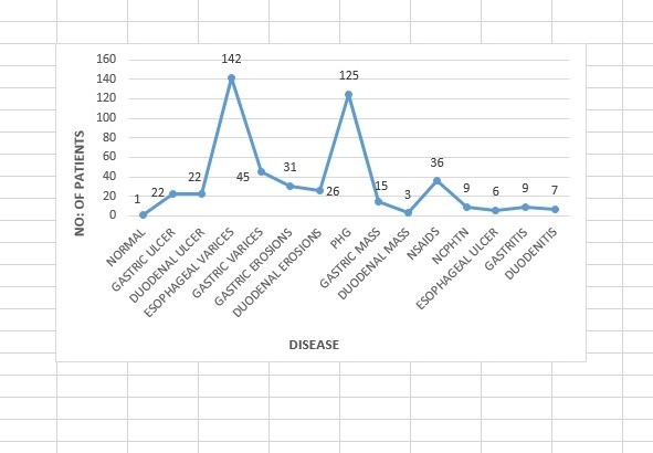
Graph 1: number of patients with different etiologies of Upper GI bleed
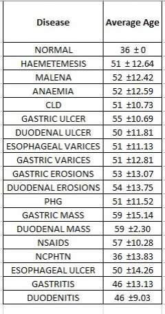
CLD was seen with highest disease frequency of 60 percent encompassing both esophageal varices as well as portal hypertensive gastropathy, as can be depicted in graph 2. Average age of patients with positive finding was in 5th decade of life as seen in table II
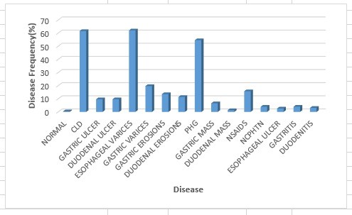
Graph 2: Disease frequency of different etiologies of upper GI bleed
Graph 2: Disease frequency of different etiologies of upper GI bleed
Upper gastrointestinal bleed, demarcated as bleeding above the ligament of Treitz in the gastrointestinal tract, presents as hematemesis, melena or hematochezia. It accounts for about 32-36% of all hospital admissions, according to various surveys done across the US and UK, 8
Literature review of other international studies done in this context also showed the same picture. Research done at Chile over three years reported that 72% of the total 249 patients had the non-variceal cause of upper GI bleed. Out of those non-variceal causes, 44% had peptic ulcer disease.9
This has created a huge difference in our study's observations compared to other studies done in this context. Our study showed 81% of cases of upper GI bleed were with variceal bleed including both esophageal and gastric varices. The only plausible explanation for this significant difference between our observation and other international studies can be an extreme predominance of viral hepatitis in our country. On one hand this significant increase in incidence of viral hepatitis has made varices the major cause of upper GI bleed. On the other hand, there is a significant fall in the incidence of peptic ulcer. Over-the-counter use of PPI over a prolonged period could have been a factor to protect against peptic ulcer disease.
Our study can be compared to many other local studies wherein varices were found to be the predominant cause of upper GI bleed. A study conducted in Jamshoro, Pakistan revealed 54% of cases as a cause of upper GI bleed.10 Another study conducted in Rawalpindi also showed esophageal varices as the most common cause of upper gastrointestinal bleed followed by duodenal ulcers.11 similarly a study conducted in Karachi revealed portal hypertension leading to varices as the most common cause of upper GI bleed.12
Among non-variceal causes of upper GI bleed, portal hypertensive gastropathy was the most common cause of upper GI bleed. We could not find such predominance in any of the studies done previously, the reason behind this could be merging of this separate entity of portal hypertensive gastropathy with other gastropathies and even considering this separate entity of portal hypertensive gastropathy a part of variceal bleed on account of having same etiology. Hence it could not have been identified as a separate entity during endoscopy and during interpretation of results.
The next most common cause among non-variceal group was NSAID induced gastritis which was found to be more common in our setup as compared to peptic ulcer disease. As far as predominance of NSAID induced gastritis over peptic ulcer disease is concerned, several other research done locally as well as across borders have shown similar results. 3,11,12,13 A study done in Nepal also showed PUD as the most common cause.7 According to another study, most common causes of upper gastrointestinal bleeding include peptic ulcer disease, varices, and esophagitis respectively. 14 A retrospective study conducted in Nigeria also revealed peptic ulcer disease as the most common endoscopic finding among patients with upper gastrointestinal bleed. 15 According to a study conducted in Turkey, the major cause of non-variceal bleeding is peptic ulcer disease, with decreased incidence of duodenal ulcers as compared to gastric ulcers, mainly due to widespread eradication of Helicobacter pylori.16 In contrast, our study showed equal presentation of gastric as well as duodenal ulcer. However, there was a predominance of gastric erosions over duodenal erosions which can be compared and attributed to previously mentioned theory of eradication of H pylori but this difference was very minimal. There was however a significant predominance of gastric mass over duodenal mass which is more common and well-established fact
Surprisingly a female predominance was observed in our study as compared to male population, with female being about 65% and males about 35% which is a significant difference. This contrasted with most of the studies done previously which have shown prominent male predominance.7,11,12,16,17,18 This significant difference in male to female presentation can’t be overlooked and need to be given due consideration. One possible reason for this can be poor concerns about female health in our resource restricted country which on one hand is leading to increase transmission of disease from male partner, while on other hand it can be due to neglected health of females over a long period of time with ultimate presentation with decompensation and upper GI bleed. Another possible reason for this can be due to the primary etiology of upper GI bleed being autoimmune hepatitis besides well-known predominance of viral hepatitis. Autoimmune hepatitis obviously has female predominance. Variable age groups at presentation with Upper GI bleed have been mentioned in different literature. In our study we reported patients in age range of 36-59 years with maximum patients presenting in 5th decade of life. The youngest age group of patients presenting with upper GI bleed was with non-cirrhotic portal hypertension presenting at a mean age of 36 years followed by gastritis and duodenitis during 4th decade of life. All remaining patients in our study were in the 5th decade of their life. Median age of patient with first three most common causes of esophageal varices, portal hypertensive gastropathy and gastric varices was 51 years. This was in accordance with a local study done previously which showed a mean age of presentation at 54.7 years. This age pattern was also observed in many other studies done previously.19, 20 However few notable studies from India and African countries including Nigeria and Uganda showed a younger mean age group of patients presenting with Upper GI bleed.17,21, 22, 23 This was in contrast to the results from, Greece17 and Canadian research that showed a relative trend towards older age group with an age of more than 64 years at presentation.24 A comparatively older age of presentation was also seen in a study done in Japan enrolling patients from three hospitals over a period of three years.25 this age group diversity in different world populations might be due to difference in approach to different treatment modalities. The late age of upper GI bleed presentations in western countries in mostly attributable to the use of NSAID in old age due to many reasons in old age which implicate the use of NSAIDS. It can also be related to patients living relatively longer life in western countries and thus enjoying a healthy life in younger age group.
As an overall impression created out of this study variceal bleed has been a major source of bleed in our setup with predominance in middle age group in female population.
There was a statistically significant difference between the mean readings of the AL measured by the IOL master and applanation ultrasound biometer, both the devices cannot be used interchangeably.
- DiGregorio AM, Alvey H. Gastrointestinal Bleeding. [Updated 2023 Jun 5]. In: Stat Pearls [Internet]. Treasure Island (FL): Stat Pearls Publishing; 2023 Jan-.Available from: https://www.ncbi.nlm.nih.gov/books/NBK537291/
- Tafen M, Stain SC. Upper gastrointestinal bleeding. Emergency General Surgery: A Practical Approach. 2019:87-102.
- Sher F, Ullah RS, Khan J, Mansoor SN, Ahmed N. Frequency of different causes of upper gastrointestinal bleeding using endoscopic procedure at a tertiary care hospital. Pakistan Armed Forces Medical Journal. 2014 Jun 30; 64(3):410-3.
- Nukala K, Srinivasan VR, Sagar RV. Clinical Presentation of Cases with Upper Gastro-Intestinal Bleeding. The Journal of the Association of Physicians of India. 2022 Apr 1; 70(4):11-2.
- Kim BS, Li BT, Engel A, Samra JS, Clarke S, Norton ID, Li AE. Diagnosis of gastrointestinal bleeding: A practical guide for clinicians. World journal of gastrointestinal pathophysiology. 2014 Nov 11; 5(4):467. https://doi.org/10.4291/wjgp.v5.i4.467
- Beg S, Ragunath K, Wyman A, Banks M, Trudgill N, Pritchard MD et al; Quality standards in upper gastrointestinal endoscopy: a position statement of the British Society of Gastroenterology (BSG) and Association of Upper Gastrointestinal Surgeons of Great Britain and Ireland (AUGIS). Gut. 2017 Nov 1; 66(11):1886-99. https://doi.org/10.1136/gutjnl-2017-314109
- Bhattarai S. Clinical profile and endoscopic findings in patients with upper gastrointestinal bleed attending a tertiary care hospital: a descriptive cross-sectional study. JNMA: Journal of the Nepal Medical Association. 2020 Jun; 58(226):409. https://doi.org/10.31729/jnma.4967
- Abougergi MS, Travis AC, Saltzman JR. The in-hospital mortality rate for upper GI hemorrhage has decreased over 2 decades in the United States: a nationwide analysis. GastrointestEndosc. 2015 Apr; 81(4):882-8.e1. https://doi.org/10.1016/j.gie.2014.09.027.
- Pinto C, Parra P, Magna J, Gajardo A, Berger Z, Montenegro C, Muñoz P. Hemorragia digestive alta variceal y no variceal: mortalidad intrahospitalaria y características clínicasen un hospital universitario (2015-2017) [Variceal and non-variceal upper gastrointestinal bleeding. Analysis of 249 hospitalized patients]. Rev Med Chil. 2020 Mar; 148(3):288-294. Spanish https://doi.org/10.4067/S0034-98872020000300288.
- Ghouri A, Kumar S, Bano S, Aslam S, Ghani MH. Endoscopic Evaluation of Upper Gastrointestinal Bleeding in Patients Presenting with Hematemesis within 24 Hours of Admission. J Liaquat Uni Med Health Sci 2016; 15(04): 174-78. https://doi.org/10.22442/jlumhs.161540488
- Ahmed J, Alam L, Shabbir K, Naqvi M, Haider E, Farooque A. ENDOSCOPIC FINDINGS IN PATIENTS PRESENTING WITH UPPER GI BLEED IN A TERTIARY CARE FACILITY. Pakistan Armed Forces Medical Journal. 2020 Feb 29(1):112. https://pafmj.org/PAFMJ/article/view/3947
- Ali SS, Butt N, Altaf HH, Abbasi A. The Etiology and Outcome of Upper Gastrointestinal Bleeding in Patients Presenting to Tertiary Care Hospital, Karachi. Pakistan Journal of Medical & Health Sciences. 2022 Sep 10; 16(07):646. https://doi.org/10.53350/pjmhs22167646
- Gupta T, Goyal S. Paradigm Shift in Etiology of Upper Gastrointestinal Bleed in Emergency Department. Journal of Renal and Hepatic Disorders. 2021 Mar 16; 5(1):14-8. https://doi.org/10.15586/jrenhep.v5i1.93
- Kamboj AK, Hoversten P, Leggett CL. Upper gastrointestinal bleeding: etiologies and management. InMayo Clinic Proceedings 2019 Apr 1 (Vol. 94, No. 4, pp. 697-703). Elsevier. https://doi.org/10.1016/j.mayocp.2019.01.022
- Jemilohun AC, Akande KO, Ngubor TD, Oku O, Ogunmola MI, Adesuyi YO, Jemilohun A, Akande K, Adesuyi Y. Endoscopic Findings in Patients with Upper Gastrointestinal Bleeding in Ogun State, Nigeria. Cureus. 2022 Mar 30; 14(3). https://doi.org/10.7759/cureus.23637
- Danış N, Tekin F, Akarca US, Ünal NG, Erdoğan EI, Akat K, Demirkoparan Ü, Karasu Z, Turan İ, Oruç N, Aydın A. Changing patterns of upper gastrointestinal bleeding over 23 years in Turkey. The Turkish Journal of Gastroenterology. 2019 Oct; 30(10):877. https://doi.org/10.5152/tjg.2019.19239
- Sotiropoulos C, Papantoniou K, Tsounis E, Diamantopoulou G, Konstantakis C, Theocharis G, Triantos C, Thomopoulos K. Acute Upper Gastrointestinal Bleeding: Less Severe Bleeding in More Frail and Older Patients, Comparison Between Two Time Periods Fifteen Years Apart. Gastroenterology Research. 2022 Jun; 15(3):127. https://doi.org/10.14740/gr1534
- Jensen DM, Eklund S, Persson T, Ahlbom H, Stuart R, Barkun AN, et al. Reassessment of rebleeding risk of Forrest IB (oozing) peptic ulcer bleeding in a large international randomized trial. Am J Gastroenterol 2017; 112(3): 441-46. https://doi.org/10.1038/ajg.2016.582
- Laine L. Upper gastrointestinal bleeding due to a peptic ulcer. N Engl J Med 2016; 374(24): 2367-76. https://doi.org/10.1056/nejmcp1514257
- Gabr MA, Tawfik MA, El-Sawy AA. Non-variceal upper gastrointestinal bleeding in cirrhotic patients in Nile Delta. Indian J Gastroenterol 2016; 35(1): 25-32. https://doi.org/10.1007/s12664-016-0622-7
- Kiringa SK, Quinlan J, Ocama P, Mutyaba I, Kagimu M. Prevalence, short term outcome and factors associated with survival in patients suffering from upper gastrointestinal bleeding in a resource limited-setting, the case of Mulago hospital in Kampala, Uganda. African health sciences. 2020 Apr 20; 20(1):426-36. https://doi.org/10.4314/ahs.v20i1.49
- Banerjee A, Bishnu S, Dhali GK. Acute upper gastrointestinal bleed: An audit of the causes and outcomes from a tertiary care center in eastern India. Indian Journal of Gastroenterology. 2019 Jun 1; 38:190-202. https://doi.org/10.1007/s12664-018-00930-7
- Yahya H, Umar H, Shekari BT, Sani K. Endoscopy for upper gastrointestinal bleeding in a tertiary hospital in Kaduna, North-West Nigeria: Experience and findings. Annals of African Medicine. 2022 Jul; 21(3):262. https://doi.org/10.4103/aam.aam_64_21
- Lu Y, Loffroy R, Lau JY, Barkun A. Multidisciplinary management strategies for acute non variceal upper gastrointestinal bleeding. Br J Surg 2014; 101(1): e34-50. https://doi.org/10.1002/bjs.9351
- Horibe M, Iwasaki E, Bazerbachi F, Kaneko T, Matsuzaki J, Minami K, Masaoka T, Hosoe N, Ogura Y, Namiki S, Hosoda Y. Horibe GI bleeding prediction score: a simple score for triage decision-making in patients with suspected upper GI bleeding. Gastrointestinal Endoscopy. 2020 Sep 1; 92(3):578-88. https://doi.org/10.1016/j.gie.2020.03.3846

An Official Publication of
Islamabad Medical & Dental College
Vol 13 No (Suppl.)
Address of Correspondence
Hareema Saeed Khan
Email:
hareemasaeed80@gmail.com
Authors’ Contribution: 1,2Conception; Literature research; manuscript design and drafting; 3,4 Critical analysis and manuscript review; 5,6Data analysis; Manuscript Editing.
Cite this article. Zada I, Dar SI, Shah FU, Khan HS, Asim M, Fatima A. Frequency of Various Aetiological Factors in Patients Presenting with Upper Gastrointestinal Bleeding. J Islamabad Med Dental Coll. 2024; 13i(Suppl.): 568-573
DOI: https://doi.org/10.35787/jimdc.
v13i(Suppl.).1084