Primary Cutaneous Mucinous Carcinoma
Nosheen Nabi1, Qudsia Ishaq2, Umer Mehmood3, Naseer Ahmed1
1Department of Histopathology, Excel Laboratory, Blue Area, Islamabad, 2Federal Medical College Islamabad, 3Department of Histopathology, University of Lahore Teaching Hospital, Lahore
Primary mucinous carcinoma of the skin is an uncommon malignancy of sweat gland origin. It is usually misdiagnosed clinically because of its benign appearance. On histological examination it should be differentiated from metastatic mucinous carcinomas arising elsewhere in the body. We present a case of a 77 years old male presenting with a swelling on face which was diagnosed clinically as a benign lesion but on histological examination it was diagnosed as mucinous carcinoma and after thorough investigations it was labelled as primary mucinous carcinoma of skin.
Keywords: Carcinoma, Cutaneous carcinoma, Mucinous carcinoma.
Primary cutaneous mucinous carcinoma (PCMC) is a rare malignant tumor of skin adnexa. Lennox et al., was the first one to discover this rare entity in 1952.1 Till now in literature, approximately 200 cases have been reported.2 This is a locally aggressive neoplasm with a tendency to recur but metastasis is rare. Most commonly mucinous carcinomas arise in the viscera and metastasize to the skin. Therefore, metastatic mucinous carcinoma should be excluded before the diagnosis of primary mucinous carcinoma.3 Wide surgical excision with or without regional lymph node dissection is considered as the treatment of choice. Distant occurs then it becomes highly resistant to both radio and chemotherapy.4
Case Report
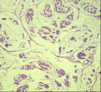
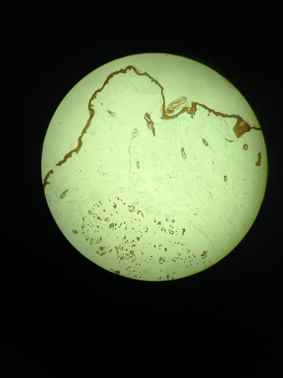
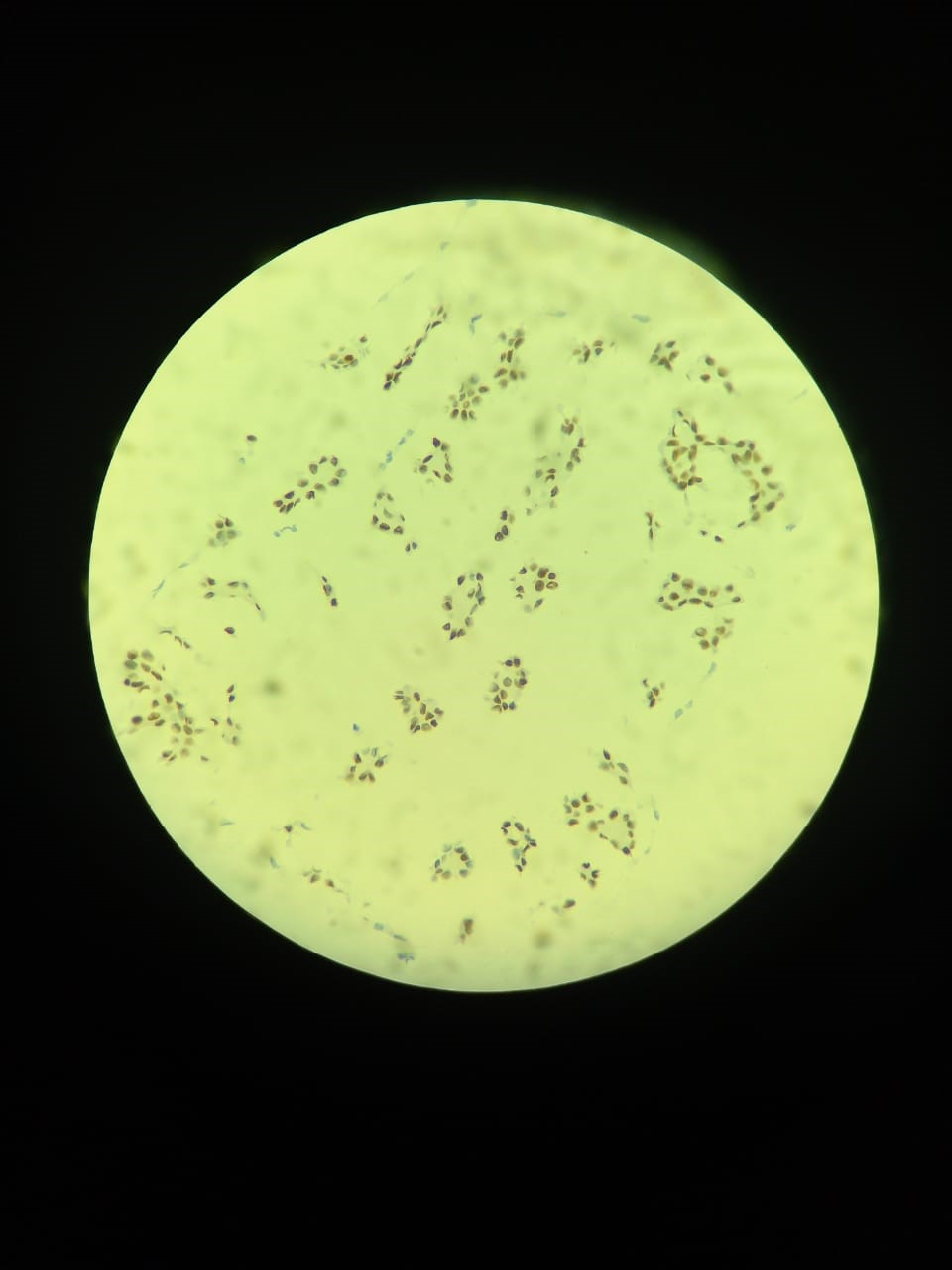
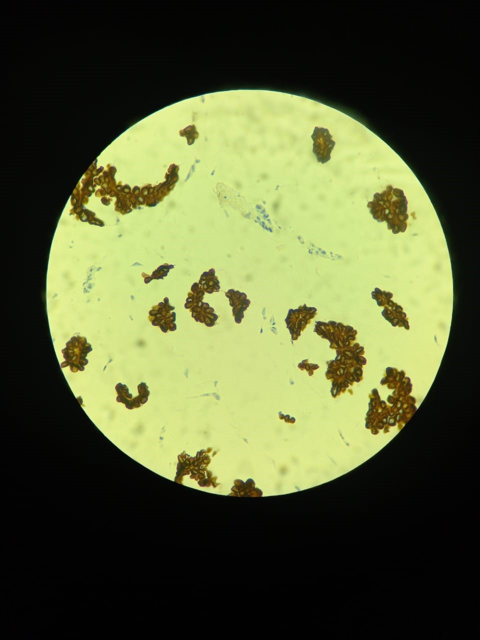
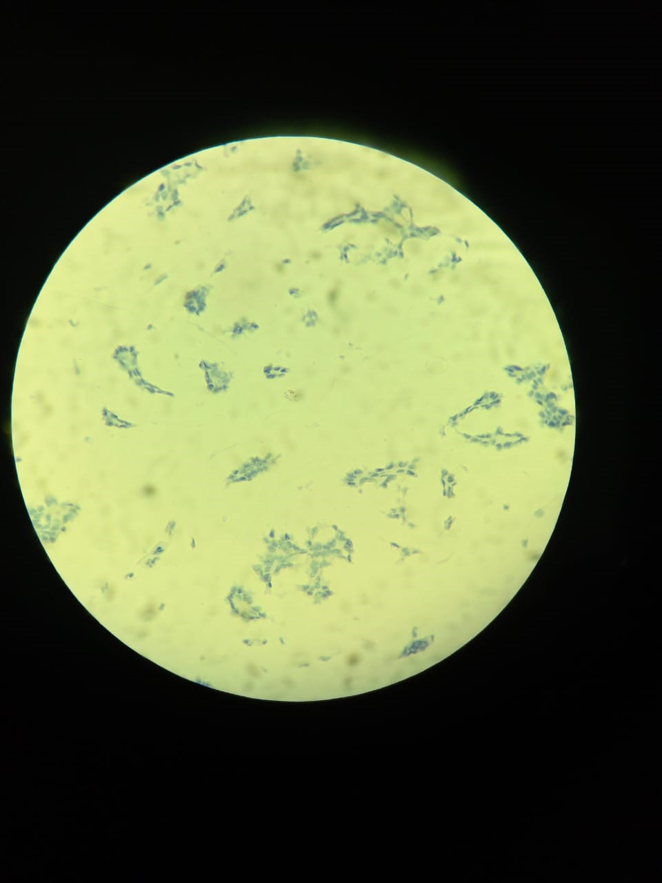
A 77 years old male was presented in a private clinic for the treatment of a boil on cheek below left eye. As it resembles a benign lesion so it was excised and no microscopic examination was done, it reappeared after some time and re excision was done not followed by histopathological examination and after some period of time it again appeared and on 3rd consecutive re excision it was sent to excel labs where its gross and histopathological examination was done. On gross examination it was a single brown soft tissue fragment measuring 0.4x0.4x0.3cm. It was entirely submitted in one cassette. On microscopic examination, the biopsy was composed of epidermis and dermis. It showed a tumor in the dermis. The tumors cells were in small clusters immersed in mucin lakes. The tumor cells had vesicular nuclei and prominent nucleoli. The lesion was involving the deep margin (Figure 1). On immuno-histochemical panel, AE/AE3 was positive, CK 20 was negative, CK7 was positive, p63 was negative, Estrogen and progesterone receptors were also positive.
A

B
C
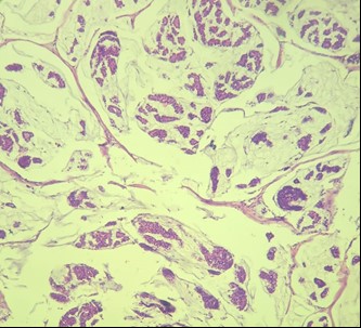
Figure 1: Morphological features of primary mucinous carcinoma of skin; A: the dermis showed nests of tumor cells in lakes of mucin (H&E X10), B: Fibrous septa around the neoplastic cells and the surrounding mucin (H&E X 20), C: The tumor cells are round to cuboid
Primary mucinous carcinoma of the skin is an uncommon malignant tumor of eccrine glands. Although it can occur at a wide age range (8-84 years), it affects individuals in 5-7th decade with approximately 2:1 male to female ratio. Increased incidence is observed in white race as compared to African American, East Asian or Indian population.5 It usually occurs in head and neck region, especially eyelids but can be found on other regions as axillae, thorax, abdomen, groin, foot, hand, and vulva. The lesion is un- encapsulated but well circumscribed, nodular, ranging in size from 0.7 to 120 cm, with the average size of around 1.8 cm. Its clinical appearance can be confused with many inflammatory, benign and malignant lesions like lipoma, epidermoid cyst, nevus, hemangioma, pyogenic granuloma, sebaceous cyst or carcinomas, metastasis from other sites, which is the most important and challenging differential because mucinous carcinomas usually metastasize to the skin from other sites.6 The most common sites from where it metastasizes are the breast, salivary glands, lacrimal glands, nose and paranasal sinuses, bronchi, renal pelvis, and ovary. This definitive diagnosis can be made by thorough clinical and radiological examination. Moreover, there are some microscopic and immunohistochemical parameters which can help differentiate primary and secondary mucinous carcinomas. In mucinous carcinomas the microscopic examination shows invasive nests of neoplastic cells arranged in glandular, cribriform or micro-papillary patterns surrounded by lakes of mucin. The individual tumor cells are usually uniform with scanty eosinophilic cytoplasm. The mucin is Periodic acid Schiff positiveand diastase resistant. This mucin is considered to be responsible for the slow growth of the tumor by depriving tumor cells from nutrients and thus rare metastasis. Primary and metastatic mucinous carcinoma shows same morphology but metastatic tumors usually show more dysplastic features which include poorly organized epithelial cells, prominent hyperchromasia and more mitotic figures than what is observed in primary mucinous carcinoma of skin.
A
B
C
D
E
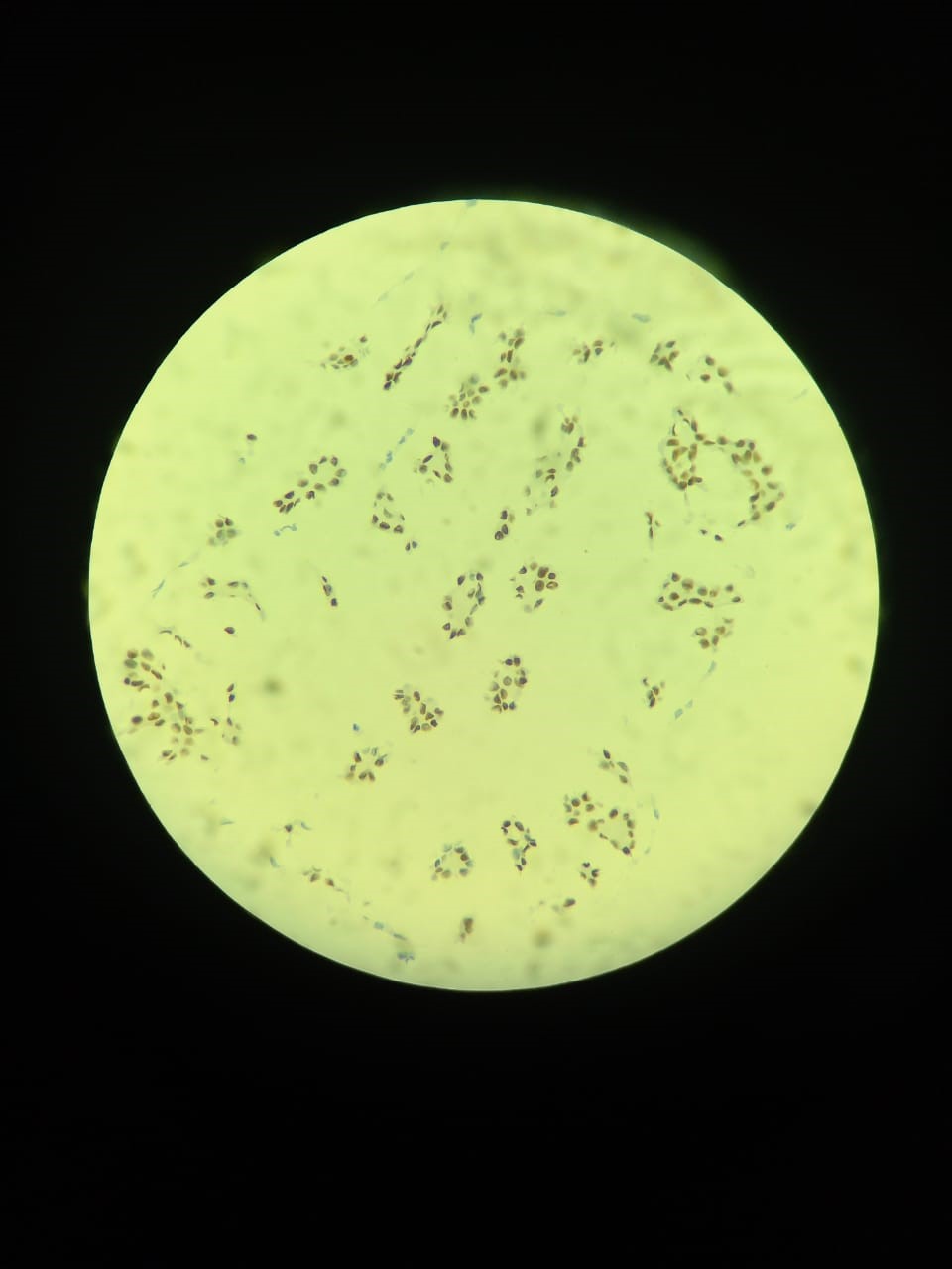
Figure 2: Immunohistochemical stains applied to PMC A: AE/AE was positive, B: CK 20 was negative, C: Ck7 was positive, D: P63 was negative, E:ER & PR was positive
The tumor usually recurs locally which is in about 30% to 40% of cases while distant metastases is rare which is approximately in 6% of cases. The prognosis of PMC is good. Older age and Asian population showed better postsurgical outcomes. Prognosis is affected by size, duration of persistence and site.10
The tumor deserves attention not only for its rarity but also for the problems of differential diagnosis with dermal metastasis from mucinous adenocarcinoma of other sites. Diagnostic concerns include its deceptively benign appearance in some cases and the difficulty in differentiating it from secondary mucinous carcinoma of skin metastasizing from a primary source elsewhere.
- Lennox B, Pearse AE, Richards HG. Mucin‐secreting tumours of the skin: With special reference to the so‐called mixed‐salivary tumour of the skin and its relation to hidradenoma. The Journal of pathology and bacteriology. 1952 Oct;64(4):865-80. https://doi.org/10.1002/path.1700640418
- Sistla R, Afroz T, Narayanswamy J. Primary mucinous adenocarcinoma of skin: myoepithelial cells are a clue to its diagnosis. Indian journal of dermatology. 2018 Mar;63(2):165 https://doi.org/10.4103%2Fijd.IJD_619_16
- Tillit SM, Iyer SS, Grieser EJ, LiVecchi JT. Treatment of Recurrent Primary Cutaneous Mucinous Carcinoma of the Eyelid with Modified Wide Local Excision. Case Reports in Ophthalmological Medicine. 2020 Dec 18;2020. https://doi.org/10.1155/2020/6668640
- Miyachi H, Hashimoto H, Suehiro K, Yamamoto Y, Ota S, Nakatani Y, Matsue H. Aromatase inhibitor for the treatment of primary mucinous carcinoma of the skin with distant metastasis. JAAD case reports. 2018;4(3):235. https://doi.org/10.1016%2Fj.jdcr.2017.10.021
- Al Beteddini OS, Sheikh S, Shareefi F, Shahab R. Primary mucinous adenocarcinoma of the scalp: a case report and literature review. International journal of surgery case reports. 2015 Jan 1;1 0:241-4. https://doi.org/10.1016%2Fj.ijscr.2015.02.006
- Kumar PA, Paulraj S, Hahn SS, Sivapiragasam A. Primary Mucinous Adenocarcinoma of the Upper Eyelid in an African American Female: A Rare Clinical Entity. A Case Report and Literature Review. Cureus. 2019 Nov;11(11). https://doi.org/10.7759/cureus.6254
- Abiola OO, Ano-Edward GH, Oluwumi OA, Lasisi ME. Primary cutaneous mucinous carcinoma of the scrotum: A rare tumor at a rare site–A case report and review of literature. Urology annals. 2020 Jan;12(1):83. https://doi.org/10.4103/ua.ua_126_18
- Brown CA, Lynch MC, Ueno CM. Mucinous adenocarcinoma of the scalp: primary cutaneous neoplasm versus underlying metastatic disease. Plastic and Reconstructive Surgery Global Open. 2018 Apr;6(4). https://doi.org/10.1097/gox.0000000000001761
- Saito Y, Ota K, Sumita Y. Primary mucinous carcinoma of the skin arising from the upper eyelid: A case report and literature review. JPRAS open. 2020 Sep 1;25:18-23. https://doi.org/10.1016%2Fj.jpra.2020.04.002
- Goyal R, Tanveer N, Singh UR. Primary mucinous eccrine adenocarcinoma: A rare skin adnexal tumor at an unusual site. Journal of cytology. 2019 Jan;36(1):71. https://doi.org/10.4103%2FJOC.JOC_28_18

An Official Publication of
Islamabad Medical & Dental College
Volume 13 Issue 1
Case Report
Address of Correspondence
Qudsia Ishaq
Email:
enayahassan00@gmail.com
Authors’ Contribution: All authors contributed equally to the conception, literature search, manuscript drafting, editing and review
Cite this article. Ishaq Q, Nabi N, Mehmood U, Ahmed N. Primary Cutaneous Mucinous Carcinoma.J Islamabad Med Dental Coll. 2024; 13(1): 183-186.
DOI: https://doi.org/10.35787/jimdc.v13i1.1088