Radiographic Assessment Regarding the Pattern of Third Molar Impaction and its Association with Gender in the Population of Islamabad
Hina Nasim1, Mawra Hyder1, Anosha Mujtaba1, Afsheen Mansoor1, Bushra Afridi1, Arsalan Hyder2
1School of Dentistry, Shaheed Zulfiqar Ali Bhutto Medical University, Islamabad; 2Department of Dentistry, Federal Government Polyclinic Hospital, Islamabad
Objective: The main objective was to determine the prevalence of impacted teeth and classify the types of impaction observed in third molars.
Methodology: This retrospective cross-sectional study spanned three months and involved the analysis of orthopantomograms (OPG) data from 278 patients, encompassing a total of 445 impacted third molars. The data were sourced from patients referred to the Radiology department of the School of Dentistry, Shaheed Zulfiqar Ali Bhutto Medical University, Islamabad, from September to December 2023. Inclusion criteria involved OPGs of individuals aged 25 years or older with fully formed roots in their third molars. Exclusion criteria included OPGs of individuals who were younger than 25 years, exhibited craniofacial anomalies, underwent prior orthodontic treatment, had erupted third molars, or presented with third molars featuring incomplete root formation.
Results: In the present study, both male and female participants were almost equal in number, and the mean age of male participants was 29.8 ± 4.4 while the mean age of female participants was 29.3 ± 4.7 Among the OPG examined, 73.2% presented with impacted mandibular third molars, while only 26.8% displayed impacted maxillary third molars. To be more precise, the left mandibular third molar was the most frequently impacted at 58.9%, whereas the left maxillary third molar had the lowest occurrence at 20.8%.
Conclusion: In conclusion, our research underscores significant gender variations in the prevalence and types of impactions observed in third molars. Additionally, a higher prevalence of mandibular third molar impaction was identified in comparison to maxillary third molars.
Keywords: Radiography, Impaction, Molar
The tooth is said to be impacted when its eruption is hampered and obstructed due to the adjacent tooth, soft tissues, inadequate space, insufficient lower jaw growth, late mineralization, hindered facial growth, and other conditions. Such teeth remain completely or partly embedded in the bone or the soft tissues, leading to pericoronitis, dental caries, and cystic lesions.1,2 As the last tooth to be erupted it faces less accessible space in the oral cavity, making eruption problematic. This is a pathological condition, in which the pathway of eruption of dentitions is obstructed and the tooth is prevented from erupting into its functional position.3
Third molars are the most impacted teeth, with a global prevalence of 30.3 to 68.6%.2,4 Another study conducted in the Gulf region suggests that the occurrence of third molar impaction is 32- 40.5 %.5 Third molars are also known as wisdom teeth since this name is associated with the time of their emergence in the oral cavity, which is early adulthood. Impaction in mandibular molars is predominantly more common than in maxillary third molars. Studies have suggested that mesioangular is a more common type of impaction found in mandibular third molars. Third molars erupt in late adolescence between the age of 18 to 20.6
According to most of the studies, there is no gender predilection, whereas some of the studies have also proposed that there is a greater incidence of impaction in females as compared to males.7 Another study reveals that males have a larger retromolar space than females of Indian heritage. The impacted third molar exhibits distinct patterns in terms of depth, position relative to the jaw, and angle relative to the occlusal plane.8 The angle of impaction is determined by using the Winters classification system, referring to the angle obtained by intersecting the longitudinal axis of the second and third molar. It is classified as mesioangular, distoangular, vertical, horizontal, and buccolingual.9
An early diagnosis is critical for its early management therefore, a detailed radiographic assessment is required to determine the pattern of the impaction, Orthopantomography (OPG) is used to measure the angulation of the impacted third molar and its relation and proximity to the underlying structures.10 Retaining the third molar might benefit orthodontic treatment, thus making it a crucial problem to be addressed.11
The objective of this study was to determine the gender-specific pattern of third molar impaction and the prevalence of third molar impaction in both arches.
This retrospective cross-sectional study spanned three months and involved the analysis of orthopantomograms (OPG) data from 278 patients, encompassing a total of 445 impacted third molars. The data were sourced from patients referred to the Radiology department of the School of Dentistry, Shaheed Zulfiqar Ali Bhutto Medical University, Islamabad, from September to December 2023. Ethical approval was obtained from the ethical review board of the School of Dentistry, SZABMU. The sample size was determined using the online calculator Rao soft, considering a population size of 1000, a 95% confidence interval (CI), and a z-value of 1.96. Inclusion criteria involved OPGs of individuals aged 25 years or older with fully formed roots in their third molars. Exclusion criteria included OPGs of individuals who were younger than 25 years, exhibited craniofacial anomalies, underwent prior orthodontic treatment, had erupted third molars, or presented with third molars featuring incomplete root formation. Additionally, patients with incomplete records on panoramic radiographs (OPG), poor-quality OPG images, or radiographs lacking the first, second, and third molars were excluded from the study.
The study evaluated the following parameters:
- The number and frequency of impacted third molars.
- The classification of third molars is based on Winters' criteria.
The analysis of the data was carried out using SPSS software (version 20). The frequency of impaction was presented as percentages. Gender predisposition towards various impaction patterns was assessed through the chi-square test, with a significance level set at p < 0.05.
In the present study, both male and female participants were almost equal in number, and the mean age of male participants was 29.8 ± 4.4 while the mean age of female participants was 29.3 ± 4.7 (table- I).
Table I: Demographic data of study participants |
|||
Gender |
Frequency (f) |
Percentage % |
Age |
Male |
138 |
49.6 |
29.8 ± 4.4 |
Female |
140 |
50.4 |
29.3 ± 4.7 |
Among the OPG examined, 73.2% presented with impacted mandibular third molars, while only 26.8% displayed impacted maxillary third molars. To be more precise, the left mandibular third molar was the most frequently impacted at 58.9%, whereas the left maxillary third molar had the lowest occurrence at 20.8% (Figure 1). Concerning the maxillary arch, the majority 48.7% showed the distoangular type of impaction, specifically in the left maxillary quadrant, the predominant pattern of third molar impaction among patients was distoangular impaction (8.6%), with only 1.4% experiencing buccolingual impaction. Likewise, in the right maxillary quadrant, the majority (9.7%) of third molars exhibited distoangular impaction, while only 1.1% displayed buccolingual impaction. Within the mandibular arch, mesioangular impaction was prevalent at 41.7%, notably in the left quadrant at 23.0%, while buccolingual impaction was the least frequent at 1.1%. Similarly, within the mandibular right quadrant, mesioangular impaction was the most common (26.3%), while buccolingual impaction had the lowest reported incidence at 0.7% Table 2.
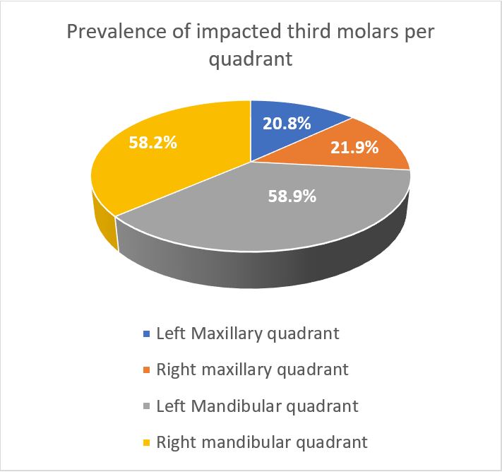
Figure 1: Prevalence of impacted third molars per quadrant among study participants
Table II: The pattern of third molar impaction per quadrant |
|||
Quadrant |
Pattern of impaction |
Frequency (f) |
Percentage % |
Left maxillary quadrant |
Mesioangular |
12 |
4.3 |
Distoangular |
24 |
8.6 |
|
Horizontal |
- |
- |
|
Vertical |
18 |
6.5 |
|
Buccolingual |
4 |
1.4 |
|
Right maxillary quadrant |
Mesioangular |
9 |
3.2 |
Distoangular |
27 |
9.7 |
|
Horizontal |
- |
- |
|
Vertical |
22 |
7.9 |
|
Buccolingual |
3 |
1.1 |
|
Left mandibular quadrant |
Mesioangular |
64 |
23.0 |
Distoangular |
28 |
10.1 |
|
Horizontal |
34 |
12.2 |
|
Vertical |
35 |
12.6 |
|
Buccolingual |
3 |
1.1 |
|
Right mandibular quadrant |
Mesioangular |
73 |
26.3 |
Distoangular |
22 |
7.9 |
|
Horizontal |
26 |
9.4 |
|
Vertical |
39 |
14.0 |
|
Buccolingual |
2 |
0.7 |
|
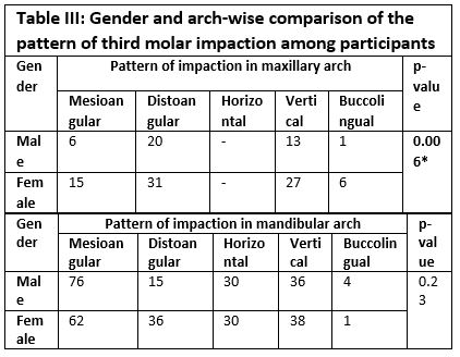
Concerning the maxillary arch, a statistically significant correlation was observed between gender and the type of impaction (p=0.006). A distinct impaction pattern was more prevalent among females compared to males. Notably, no horizontal impaction pattern was observed in either gender. Conversely, in the mandibular arch, no statistically significant association was identified between gender and type of impaction (p=0.23) (refer to Table 3). Additionally, a slightly higher incidence of mesioangular (47.2%) was noted in males, while females exhibited a higher prevalence of distoangular impactions (37.1%).
The present study aimed to determine the prevalence of impacted teeth and classify the types of impaction observed in third molars. Among the examined cases 73.2% exhibited impaction in the mandibular third molar, with the remaining 26.8% demonstrating impaction in the maxillary third molars. Similarly, a study conducted in Malaysia revealed a higher prevalence of impaction in mandibular third molars (52.2%) compared to maxillary third molars (47.7%).3 Likewise, a study carried out in India indicated a higher prevalence of impacted mandibular third molars (57.9%) in comparison to impacted maxillary third molars (42%).13 Also, a study conducted in Oman revealed a higher incidence of impacted third mandibular molars, with 56%, compared to impacted third maxillary molars, which accounted for 44%.14
The current investigation indicated that the prevailing impaction pattern for third molars in the maxillary arch was distoangular, while in the mandibular arch, it was mesioangular. Likewise, a study conducted in Saudi Arabia also found that the distoangular impaction pattern was the most prevalent type in the maxillary arch, accounting for 96.6%.15 Another study conducted in India revealed that vertical impaction (62.5%) and distoangular impaction (33.1%) are the most frequently observed types of third molar impaction in the maxillary arch.16 Similarly, a study undertaken in Kuwait found that the majority of impactions of third molars in the maxillary arch were of the distoangular type, comprising 68.5%.17 A study carried out in Iran documented that the prevalent form of impaction for mandibular third molars was mesioangular, accounting for 35.9%11 In another study conducted in Lebanon, it was reported that the majority (46.5%) of mandibular third molars exhibited a mesioangular pattern of impaction.18 Likewise, in a study conducted in Estonia, it was observed that the mesioangular pattern of impaction was the most commonly encountered type among mandibular third molar impactions 52.5%.19
This study found a significant correlation between gender and the impaction pattern of maxillary third molars, but no such association for mandibular third molars. Among females in the maxillary arch, various impaction types were prevalent, including mesioangular, distoangular, vertical, and buccolingual, while males predominantly exhibited mesioangular impaction for mandibular third molars compared to females. Similarly, a study conducted in India reported the majority of males (59.3%) presented with mesioangular type of mandibular third molar impaction as compared to females (40.6%).20 Likewise, a study conducted in India indicated that males (25.08%) exhibit a higher tendency for the mesioangular pattern of impaction in mandibular third molars as compared to females (17.08%).21 In contrast to our findings, a study conducted in Nigeria revealed that females exhibited a higher prevalence of mesioangular mandibular third molar impaction (68%) compared to males (44.4%).1 Unlike our study, a previous study conducted in Saudi Arabia showed no sex predilection for maxillary third molar impaction, which shows females are more prone to have maxillary impactions as compared to males.22 The evaluation of OPGs was carried out within a limited timeframe; therefore, a more comprehensive sample size might provide more conclusive insights into our observations. Due to the study's single-centre design, it is essential to approach generalizations of the data with caution. Consequently, conducting multi-centred research studies with larger sample sizes becomes imperative to support and confirm our findings.
In conclusion, our research underscores significant gender variations in the prevalence and types of impactions observed in third molars. Additionally, a higher prevalence of mandibular third molar impaction was identified in comparison to maxillary third molars. The predominant impaction pattern in the mandibular arch was mesioangular, contrasting with the distoangular impaction predominance in the maxillary arch. These insights offer valuable information for clinicians, emphasizing the importance of considering gender-specific factors when assessing and managing third molar impactions in both arches.
- Obuekwe O, Enabulele J. Gender variation in pattern of mandibular third molar impaction. Age (years). J Dent Health Oral Disord Ther 2017;20(5):2-8. https://doi.org/org/10.15226/jdodt.2017.00177
- Kumar SM, Al-Hobeira H, Shaikh S, Siddiqui AA, Syed J, Mian R. Distribution of impacted third molars based on gender and patterns of angulation in dental students of the Hai’l region, Saudi Arabia: A panoramic radiographic (OPG) Study. Int J Contemp Med Res. 2017;4(9):1829-32.
- Ahmad P, V'vian T, Chaudhary F, Chaudhary A, Haseeb A, Yaqoob M, et al. Pattern of third molar impactions in north-eastern peninsular Malaysia: A 10-year retrospective study. Niger J Clin Pract 2021;24(7):1028-36. https://doi.org/ 10.4103/njcp.njcp_499_20
- Tenrilili ANA, Yunus B, Rahman FUA. Third molar impaction prevalence and pattern: a panoramic radiography investigation. Jurnal Radiologi Dentomaksilofasial Indonesia (JRDI). 2023;7(1):9-14. https://doi.org/10.32793/jrdi.v7i1.951
- Al-Anqudi SM, Al-Sudairy S, Al-Hosni A, Al-Maniri A. Prevalence and pattern of third molar impaction: a retrospective study of radiographs in Oman. Sultan Qaboos Univ Med J 2014;14(3):e388.
- Rauf S, Ali W, Tariq Q, Chaudhry R, Kazmi SS, Imtiaz M. Pattern of mandibular third molar impaction: a radiographic study. Pak Oral Dent J 2 2019;39(3):238-42.
- Hasan KM, Sobhana C, Rawat SK, Singh D, Mongia P, Fakhruddin A. Third molar impaction in different facial types and mandibular length: A cross-sectional study. Natl j maxillofac surg. 2021;12(1):83. https://doi.org/10.4103/njms.NJMS_111_20
- Salam S, Bary A, Sayed A. Prevalence of Impacted Teeth and Pattern of Third Molar Impaction among Kerala Population a Cross Sectional Study. J Pharm Bioallied Sci 2023;15(Suppl 1):S354-S7. https://doi.org/10.4103/jpbs.jpbs_618_22
- Carter K, Worthington S. Predictors of third molar impaction: a systematic review and meta-analysis. J Dent Res. 2016;95(3):267-76. https://doi.org/10.1177/0022034515615857
- Shareif MS, Paul S, Ghani NFC, Fareez IM. Pattern of mandibular third molar impaction in Malaysia population and their association with gender, age and race. J Int Dent Medical Res. 2020;13(1):194-200.
- Rezaei F, Imani MM, Khavid A, Nabavi A. Patterns of mandibular third molar impaction in an Iranian subpopulation. Pesqui. Bras Odontopediatria Clín Integr 2020;20. https://doi.org/10.1590/pboci.2020.099
- Jaroń A, Trybek G. The Pattern of Mandibular Third Molar Impaction and Assessment of Surgery Difficulty: A Retrospective Study of Radiographs in East Baltic Population. Int J Environ Res Public Health. 2021;18(11). https://doi.org/10.3390/ijerph18116016
- Jain S, Debbarma S, Prasad SS. Prevalence of Impacted Third Molars among Orthodontic Patients in Different Malocclusions. Indian J Dent Res. 2019;30(2):238-42. https://doi.org/10.4103/ijdr.IJDR_62_17
- Sayed N, Bakathir A, Pasha M, Al-Sudairy S. Complications of Third Molar Extraction: A retrospective study from a tertiary healthcare centre in Oman. Sultan Qaboos Univ Med. 2019;19(3):e230-e5. https://doi.org/10.18295/squmj.2019.19.03.009
- Qassadi TM, Shafei AA, Alhazmi AA, Odabi NI. Prevalence and pattern of third molar impaction among the Saudi Population in Jazan Region, Saudi Arabia. Saudi J Oral Dent Res. 2020;5:36-42.
- Rupakar P, Maheswari J, Yadav S, Dudhat D, Shah K, Patel S. Assessment of Third Molar Impaction Pattern in the Mandible and in the Maxilla. Indian J Public Health Res Dev. 2022;13(3). https://doi.org/10.37506/ijphrd.v13i3.18215
- Ali D. Pattern of Third Molar Impactions in the Kuwaiti Population: Retrospective Radiographic Study. J Int Dent Medical Res. 2020;13(4):1494-8.
- Khouri C, Aoun G, Khouri C, Saade M, Salameh Z, Berberi A. Evaluation of third molar impaction distribution and patterns in a sample of Lebanese population. J Maxillofac Oral Surg 2022:1-9. https://doi.org/10.1007/s12663-020-01415-x
- Jaroń A, Trybek G. The pattern of mandibular third molar impaction and assessment of surgery difficulty: A Retrospective study of radiographs in east Baltic population. Int J Environ Res Public Health. 2021;18(11):6016. https://doi.org/ 10.3390/ijerph18116016
- Passi D, Singh G, Dutta S, Srivastava D, Chandra L, Mishra S, et al. Study of pattern and prevalence of mandibular impacted third molar among Delhi-National Capital Region population with newer proposed classification of mandibular impacted third molar: A retrospective study. Natl J Maxillofac Surg. 2019;10(1):59-67. https://doi.org/ 10.4103/njms.NJMS_70_17
- Retrospective Study of Pattern of Mandibular Third Molar Impaction in South Indian Population. I ndian J Forensic Med Toxicol. 2020;14(4):6120-30. https://doi.org/10.37506/ijfmt.v14i4.12558
- Alfadil L, Almajed E. Prevalence of impacted third molars and the reason for extraction in Saudi Arabia. Saudi Dent J. 2020;32(5):262-8. https://doi.org/10.1016/j.sdentj.2020.01.002

An Official Publication of
Islamabad Medical & Dental College
Volume 13 Issue 1
Address of Correspondence
Hina Nasim
Email:
drhinanasim1990@gmail.com
Authors’ Contribution: 1,2Conception; Literature research; manuscript design and drafting; 3,4 Critical analysis and manuscript review; 5,6Data analysis; Manuscript Editing.
Cite this article. Nasim H, Hyder M, Mujtaba A, Mansoor A, Afridi B, Hyder A. Radiographic Assessment Regarding the Pattern of Third Molar Impaction and its Association with Gender in the Population of Islamabad. J Islamabad Med Dental Coll. 2024; 13(1): 54-59.
DOI: https://doi.org/10.35787/jimdc.v13i1.1123