Applanation Ultrasound Versus Optical Biometry in the Measurement of Axial Length in a Normal Eye
Arsh Zahoor, Muhammad Azam Khan, Irfan Aslam Khattak, Ayisha Shakeel, Maria Saleem, Shaista Haider
Department of Ophthalmology, Pakistan Air Force Hospital, Islamabad
Objective: Accurate measurement of the axial length (AL) of the eye is important to make sure that the patient is emmetropic postoperatively. Multiple studies conducted around the globe compare AL using optical and ultrasound biometry. However, results are still inconclusive, as some studies stated that there is no difference in both modalities, whereas other studies considered optical biometry to be more superior. We aim to compare AL measurements using applanation ultrasound and optical biometry AL Scan, in Islamabad, in order to find out which modality is more superior.
Methodology: This was a prospective cross-sectional study that was conducted at the Department of Ophthalmology, PAF Hospital, Islamabad. The study was conducted over a period of eight months, from April to November 2023. Healthy participants with normal anterior and posterior segments were enrolled using non-probability, consecutive sampling. The axial length of each eye was measured by both optical and ultrasound biometer.
Results: There were 80 eyes of 40 participants including both males (77.5%) and females (22.5%) aged between 16-58 years. The axial length using the optical biometer showed a greater value ranging from 22.01 to 30.18mm with a mean value of 23.89 ± 1.31mm whereas the axial length measured using applanation ultrasound was 21.62 to 30.00mm with a mean of 23.53 ± 1.29mm. The mean difference between both was 0.36 ± 0.21mm which was statistically significant.
Conclusion: There is a statistically significant difference between the mean readings of the AL measured by the optical biometer and ultrasound biometer, so both the devices cannot be used interchangeably.
Keywords: Biometry, cataract, lenses, intraocular
The incidence of cataract is high, and it causes significant visual disturbance. It that can only be definitely treated by surgical procedures in which the physiological lens is replaced by an artificial lens1,2 The process of calculating the power of the IOL to be implanted is known as biometry and it is performed by using mathematical formulas that require biometric readings of the eye.2 Axial length (AL), which refers to the length of the eye from the anterior surface of the cornea to the internal limiting membrane (ILM) of the retina or retinal pigment epithelium (RPE), is a reading required in all power calculation formulas and can be measured by ultrasound or optical biometry before surgery.[3] AL is an important indicator of the refractive status of the eye, with an average value of 23.1±1.1 mm (mean±SD).4 Shorter lengths (<22 mm) are usually found in patients with hyperopia and longer lengths (> 26 mm) are typically found in myopic patients.[3] Therefore, accurate measurements of the AL is important as an underestimation or overestimation of the AL can end up causing a myopic or hyperopic shift respectively, postoperatively.5,6 Different technologies have been designed and introduced in order to allow accurate and precise biometric measurements of the eye.1 AL measurement in optical biometry is based on non-contact partial coherence laser interferometry (signals of retinal pigment epithelium) principle while in acoustic biometry is based on A scan ultrasound method (signals from internal limiting membrane).7 Ultrasound (US) biometry is the gold standard method of Intraocular Lens (IOL) power calculation but the corneal indentation with the probe underestimates the axial length.6 It also typically requires more time, human personnel power, extra training and a higher level of patient contact.1 These drawbacks are overcome by the newer optical biometry devices which uses light to measure the ocular distances.6 Optical biometry is one of the main procedures that is being performed over a long period of time.1 It has some advantages, since it prevents the risk of transmitting infection, corneal abrasion, and done without topical anesthesia. However, it requires patient co-operation and, more importantly, optical biometry cannot be performed accurately in the presence of mature cataracts, posterior subcapsular cataracts, maculopathy, or retinal pathologies.6,8 In these situations, ultrasound biometry often is the only tool available. Measurement of AL is important in managing certain ocular conditions and more importantly for the determination of the power of the IOL to be implanted after cataract surgery. Therefore, we need to have access to devices that can be used interchangeably and have good repeatability. If this is achieved, then we can use optical devices with full confidence and prevent transmission of infection from one patient to another, as is the drawback for ultrasound. Multiple studies conducted around the globe compare AL using optical and ultrasound biometry. However, results are conflicting as some studies stated that there is no difference in both modalities1, whereas other studies considered optical biometry to be more superior.6 We aim to compare AL measurements using applanation ultrasound and optical biometry AL Scan, in Islamabad, in order to find out which modality is more superior.
This was a prospective cross-sectional study that was conducted at the department of Ophthalmology, PAF Hospital Islamabad. The study was conducted over a period of eight months, from April to November 2023, after the approval of the ethical committee. Calculation of the sample size was done with the help of WHO calculator with a confidence interval of 95% and 0.05 margin of error. 40 participants (80 eyes) were enrolled after taking written consent. Non-probability, consecutive sampling was used. Participants were of both the genders, aged 15-60 years, and included healthy hospital staff members, healthy volunteers such as patients’ attendants, and candidates presenting to our OPD for medical fitness with normal eyes. Patients with refractive errors corrected to 6/6 with spectacles were also included in the study. Participants younger than 15 and older than 60 years of age were excluded from the study, as were those who had history of any ocular surgery or trauma. Patients with refractive errors not corrected to 6/6 were also excluded. Presence of any anterior or posterior segment pathology including cataract, the use of any topical medication and uncooperative patients with physical or mental limitations were also not included.
Axial length was measured by the optical biometer (NIDEK Optical Biometer AL Scan) first. Subjects were asked to sit with their chin up and their forehead touching the forehead bar, lateral lid canthus was aligned with the engraved lines on the device. They were asked to look at the fixation target. Multiple images were captured by the device and it measured the AL. Three consecutive readings were taken and an average AL was recorded. Contact biometry was performed afterwards in order to prevent any change in the measurement resulting from corneal aberration. Hi-Scan ultrasound 2000 by Optikon was used. Topical anesthesia (proparacaine hydrochloride 0.5%) was instilled in both eyes. After 60 seconds the subjects were told to look at a far target, the ultrasound probe was positioned right at the center of the cornea, AL was measured 3 times and an average was taken. The probe was then sterilized to avoid transmission of infection. Optical and ultrasound AL measurements were taken by different personnel in order to avoid bias.
All the data was then recorded using a structured questionnaire. The collected data was entered and then analyzed using SPSS version 26.0. All the quantitative variables, such as age and AL were shown as mean and SD. whereas, frequency and percentage were used to show qualitative variables like gender and laterality. Mean AL was compared by paired sample t-test. A p value ≤ 0.05 was considered significant. Pearson correlation was also estimated.
In this study we examined 80 eyes including both males (77.5%) and females (22.5%) (Table I). Age ranged between 16-58 years with a mean age of 29.15 ± 10.39 years (Table II). The axial length of each eye was measured using the optical biometer first which showed a greater value ranging from 22.01 to 30.18mm with a mean value of 23.89 ± 1.31mm whereas the axial length measured using applanation ultrasound was 21.62 to 30.00mm with a mean of 23.53 ± 1.29mm (Table II).
The mean difference between both was 0.36 ± 0.21mm which was found to be statistically significant (P <0.005). There was also a strong positive correlation found between both the methods (0.988) (Figure 1).
Table I: Showing the Percentage Distribution of Gender and the Eye |
||||
Frequency |
Percentage |
Cumulative Percentage |
||
Gender |
Male |
62 |
77.5 |
77.5 |
Female |
18 |
22.5 |
100 |
|
Laterality |
Right |
40 |
50 |
50 |
Left |
40 |
50 |
100 |
|
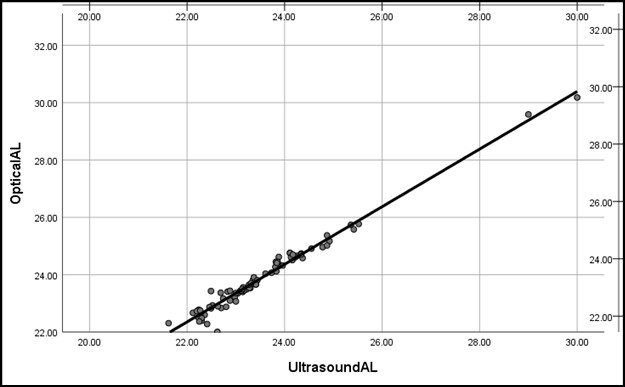
Figure 1: Correlation curve showing a statistically significant correlation
Table II: Showing the frequency description of Age and Axial Length |
||||||
|
Total |
Range |
Minimum |
Maximum |
Mean |
Std. Deviation |
Age |
80 |
42 |
16 |
58 |
29.15 |
10.394 |
Optical AL |
80 |
8.17 |
22.01 |
30.18 |
23.8879 |
1.31427 |
Ultrasound AL |
80 |
8.38 |
21.62 |
30.00 |
23.5260 |
1.29252 |
Throughout the world, different studies conducted have revealed different results and conclusions. These differences may be due to the use of devices of different generations or manufacturers, or by the difference in study populations and age rages. The expertise of the device operator may also play a role in this difference. A study carried out by the Cooke et al included 1970 eyes and the axial length was measured with both optical and ultrasonic biometer. The mean AL measured using optical biometry was only 0.0873 mm longer than that measured using ultrasound.9 Our study, however, found a difference of 0.36±0.21mm (P<0.005). This difference may have occurred because we had used contact ultrasound while they had used the immersion ultrasound device. Similarly, the results from a study conducted in India also showed comparable results for the measurement of axial length using both the optical and ultrasonic biometry with a mean difference of 0.11 ± 0.02 mm. In contrast to our study, there was no clinically significant difference (P=0.19) between the results.10 The Cho et al. study conducted in South Korea measured the axial length of patients using 6 different devices. Optical as well as ultrasonic devices were compared. From their study they had also concluded that the readings of the devices weren’t very different and either of the devices could be used with comparable results.11 In a Saudi Arabian study, the investigator measured the AL of 68 eyes using optical and ultrasonic device. They found a positive correlation r = 0.987 as well.12 In another study conducted in Iran, the biometric parameters were measured using IOLMaster700 optical biometer and US-4000 ultrasound biometer. Results also revealed a positive correlation between the readings and the devices were considered to be interchangeable.13 A study conducted in Egypt showed that there was no statistically significant difference (P <0.101) in the axial length when measured using optical biometry or ultrasonic biometry. The mean AL in the optical group in the study was 28.5 ± 1.83 while in the ultrasonic group it was 29.6 ± 2.1.14 A similar study carried out in Pakistan showed that there was a statistically significant difference (p < 0.05) while comparing non-contact and ultrasound technique in axial length measurement. The measured axial length was 23.31 ± 0.88mm that was found to be longer while using a non-contact biometer as compared to an ultrasound biometer which gave a mean axial length measurement of 22.79 ± 0.92mm. Therefore, there was a difference of 0.53 ± 0.32mm in the AL measured between both the instruments.15 Another Egyptian study showed that the measurements of the IOL calculated using the optical Zeiss IOL Master produced the best IOL power prediction leading to better post cataract surgery refractive status as compared to those with ultrasonic biometry.16 Pereira et all concluded that ultrasonic measurements of the axial length were found to be shorter than that measured by optical biometry. The postoperative refractive status was also more accurate in the group of patients with axial length measured using optical biometry.17 Our study also showed similar results with the axial length being shorter when measured with ultrasound biometer. A study conducted by Saha et al. Was also aimed at determining the difference between optical and ultrasound-based machines used for axial length measurement, but was more focused on patients with extreme myopia. Their results showed a considerably more accurate axial length measurements using optical biometry as compared with the traditional ultrasonic biometry.18 A prospective comparative study that was performed on 200 patients suggested that the AL-Scan biometer could be used for routine clinical practice to acquire accurate axial length measurements.19
The results of this study show that as there is a statistically significant difference between the mean readings of the AL measured by the optical biometer and applanation ultrasound biometer, both the devices cannot be used interchangeably. Repeated readings were taken using both devices making this a reliable study. However, this study is not free of limitations, notably due to performing this study on only two devices so the results may differ if other devices are used. We concluded that optical biometry should be preferred over ultrasound biometry in the estimation of axial length.
- Chia TMT, Nguyen MT, Jung HC. Comparison of optical biometry versus ultrasound biometry in cases with borderline signal-to-noise ratio. Clin Ophthalmol Auckl NZ. 2018;12:1757–62. https://doi.org/10.2147/opth.s170301
- Hashemi H, Miraftab M, Panahi P, Asgari S. Biometry and intraocular power calculation using a swept-source optical coherence tomography: A repeatability and agreement study. Indian J Ophthalmol. 2022;70(8):2845. https://doi.org/10.4103/ijo.ijo_249_22
- Wiseman SJ, Tatham AJ, Meijboom R, Terrera GM, Hamid C, Doubal FN, et al. Measuring axial length of the eye from magnetic resonance brain imaging. BMC Ophthalmol. 2022;22(1):54. https://doi.org/10.1186/s12886-022-02289-y
- Bikbov MM, Kazakbaeva GM, Rakhimova EM, Rusakova IA, Fakhretdinova AA, Tuliakova AM, et al. Axial length and its associations in the Ural Very Old Study. Sci Rep. 2021;11(1):18459. https://doi.org/10.1038%2Fs41598-021-98039-z
- Liu R, Li H, Li Q. Differences in Axial Length and IOL Power Based on Alternative A-Scan or Fellow-Eye Biometry in Macula-Off Rhegmatogenous Retinal Detachment Eyes. Ophthalmol Ther. 2022;11(1):347–54. https://doi.org/10.1007%2Fs40123-021-00439-x
- Mohanty G, Rajguru H, Agarwal K. Comparison of Intraocular Lens Power Estimation by Optical Biometry and Ultrasound Biometry in Cataract Surgery. J Clin Diagn Res. 2022;16(6):15–8. https://doi.org/10.7860/JCDR/2022/54834.16477
- Anwar F, Khan AA, Majhu TM, Javaid RMM, Bokhari MH, Ghaffar MT. Comparison of Accuracy of IOL Master and a Scan Acoustic Biometry in Silicon Oil Filled Eyes. Pak J Med Health Sci. 2022;16(5):267–9. https://doi.org/10.53350/pjmhs22165267
- González-Godínez S, Saucedo-Urdapilleta R, Mayorquín-Ruiz M, et al. Ocular biometry in dense cataracts: Comparison of partial-coherence interferometry, swept-source optical coherence tomography and immersion ultrasound. Indian J Ophthalmol. 2022;70(1):107-111. https://doi.org/10.2147/opth.s170301
- Cooke DL, Waldron R, Savini G, Riaz KM, Taroni L, Murphy DA, et al. Immersion ultrasound biometry vs optical biometry. J Cataract Refract Surg. 2022;48(7):819–25. https://doi.org/10.1097/j.jcrs.0000000000000865
- Paul M. Comparison of applanation ultrasound biometry with optical biometry for intraocular lens power estimation in cataract surgery and their impact on prediction error. Kerala J Ophthalmol. 2021;33(2):173. https://doi.org/ 10.4103/kjo.kjo_48_21
- Cho YJ, Lim TH, Choi KY, Cho BJ. Comparison of Ocular Biometry Using New Swept-source Optical Coherence Tomography-based Optical Biometer with Other Devices. Korean J Ophthalmol. 2018;32(4):257. https://doi.org/10.3341/kjo.2017.0091
- Nakhli FR. Comparison of optical biometry and applanation ultrasound measurements of the axial length of the eye. Saudi J Ophthalmol. 2014;28(4):287–91. https://doi.org/10.1016/j.sjopt.2014.04.003
- Khorrami-Nejad M, Khodair AM, Khodaparast M, Babapour Mofrad F, Dehghanian Nasrabadi F. Comparison of the ocular ultrasonic and optical biometry devices in the different quality measurements. Journal of Optometry. 2023;16(4):284–95. https://doi.org/10.1016%2Fj.optom.2023.05.001
- El Enin MAA, Mohamed AR, Fayad AESEG. Optical Biometry versus Ultrasonic Biometry in Intraocular Lens Power Calculations in High Axial Myopia. Al-Azhar International Medical Journal [Internet]. 2023;4(2). Available from: https://aimj.researchcommons.org/journal/vol4/iss2/20 https://doi.org/10.58675/2682-339X.1675
- Ashraf MA, Sarwar MS, Afzal MA, Khalid I, Shahid S. Comparison of Axial Ocular Measurements with Contact and Non-contact Biometry. Pak J Ophthalmol. 2020 [cited 2024;36(1).
- Elsaadani AEI, Badawi NM, Elterawy AZA. Optical Biometry Versus Ultrasound Biometry. The Egyptian Journal of Hospital Medicine. 2020;81(5):2088–92. https://dx.doi.org/10.21608/ejhm.2020.126408
- Pereira A, Popovic M, Lloyd JC, El-Defrawy S, Schlenker MB. Preoperative measurements for cataract surgery: a comparison of ultrasound and optical biometric devices. Int Ophthalmol. 2021;41(4):1521–30. https://doi.org/10.1007/s10792-021-01714-3
- Saha R, Sinharay A, Kuila J, Ganguly P. I.O.L. Master 700 Optical Biometry versus Conventional Ultrasound Biometry (Immersion Technique) in Intra Ocular Lens Power Calculation in High Myopia Patients. jebmh. 2020;7(23):1088–91. http://dx.doi.org/10.18410/jebmh/2020/235
- Kumari A, Sinha S. To evaluate the clinical accuracy of ultrasound and optical biometry: a comparative study. IJMBS [Internet]. 2020 May 30 [cited 2024 Mar 3];4(5). Available from: https://ijmbs.info/index.php/ijmbs/article/view/1199. https://doi.org/10.1097/j.jcrs.0000000000000865

An Official Publication of
Islamabad Medical & Dental College
Volume 13 Issue 2
Address of Correspondence
Arsh Zahoor
Email:
arshkj_zahoor@gmail.com
Authors’ Contribution: 1,2Conception; Literature research; manuscript design and drafting; 3,4 Critical analysis and manuscript review; 5,6Data analysis; Manuscript Editing.
Cite this article. Zahoor A, Khan MA, Khattak IA, Shakeel A, Saleem M, Haider S. Applanation Ultrasound Versus Optical Biometry in the Measurement of Axial Length in a Normal Eye. J Islamabad Med Dental Coll. 2024; 13(2): 300-305.
DOI: https://doi.org/10.35787/jimdc.v13i2.1164