Comparison of Clinical, Etiological and Antibiotic Sensitivity Patterns of UTI in CAKUT and Non CAKUT Children
Nehaj Tariq1, Shahina Yasmin2, Tariq Butt2, Aleena Butt2, Shazia Arif, Saeeda Bano2
1Department of Microbiology, Watim Medical and Dental College, Islamabad; 2Department of Microbiology, Islamic International Medical College Trust, Rawalpindi; 3Department of Microbiology, NUST School of Health Sciences
Objective: The current study was conducted to evaluate the accuracy of already in use phenotypic methods of identification of MRSA, keeping in view the gold standard method of mec-A gene detection using Polymerase chain reaction (PCR) and to compare the sensitivity and specificity of different phenotypic methods with the genotypic method (PCR).
Methodology: A descriptive cross-sectional study was carried out at Microbiology Lab, Pakistan Railway Hospital, Rawalpindi from October 2021 to September 2022, after approval from Institutional Ethical Review Committee (Riphah/IIMC/IRC/21/73), using non-probability sampling technique. A total of 222 samples of Staphylococcus aureus were isolated from all the clinical specimens received at Microbiology lab at Pakistan Railway Hospital. Among those isolates, 150(67.5%) were Methicillin sensitive Staphylococcus aureus (MSSA) and 72(32.4%) were Methicillin Resistant Staphylococcus aureus (MRSA). The results of Phenotypic methods were compared with the results of PCR by using Chi-square formula.
Results: The sensitivity and specificity of Cefoxitin Disc Diffusion method was 62.5% and 96.6%, Oxacillin Disc Diffusion method was 65.3% and 93.3%, Oxacillin Screen Agar technique was 81.9% and 96% while that of Oxacillin MIC by Agar Dilution was 91.6% and 89.3% respectively. Oxacillin screen agar demonstrates the better phenotypic technique for detection of MRSA in routine practice.
Keywords: Cefoxitin Disc Diffusion, Oxacillin Disk Diffusion, Oxacillin Screen Agar, Oxacillin MIC, PCR.
Staphylococcus aureus is one of the most common bacteria isolated in the clinical laboratory. It is a gram-positive bacterium and an important member of the normal flora.1 It lives as a colonizer under normal circumstances but can cause several health issues when becomes invasive. In spite of hygienic improvements and usage of effective antimicrobials; it is an important contributor in both hospital and community infectious load.2 Staphylococcus aureus produces a variety of enzymes and toxins making it a highly virulent organism.3 It’s invasiveness due to the surface proteins, endospore and biofilm formation, leads to recurrent infections.4 Methicillin-resistant Staphylococcus aureus (MRSA); a clinical variant has raised serious health concerns because of rapid transmission and spread, causing outbreaks in the clinical setups. Multi drug resistance (MDR) has become a major concern mainly due to the empirical antimicrobial drugs usage. Therefore administration of more expensive and tiresome management is required.5 Methicillin/oxacillin-resistant Staphylococcus aureus are heterogeneous in their expression of resistance to β-lactam agents. Accurate identification of MRSA is very important for enhancing the reliability of the patient management.6 β-lactams, are ineffective against MRSA strains except for some new cephalosporins e.g., Ceftaroline.7 The mecA gene present at the staphylococcal cassette chromosome (SCC) is responsible for methicillin or oxacillin resistance and for resistance to nafcillin too; therefore, the resistance is independent of ß-lactamase production. There are 11 types based on the combination of mecA and ccr complexes. Hospital acquired-MRSA isolates have SCCmec types I, II, or III, while the routine community acquired-MRSA strains are SCC mec type IV or V, 8 mec A gene encodes an additional penicillin binding protein (PBP)-2a or PBP2’ which has a low affinity for β lactam antibiotics. The gold standard method for identification of mec-A gene is PCR, but this facility is neither available nor economical for routine purpose in most of the clinical laboratories.9 Therefore, one of the available phenotypic methods is adopted in routine laboratory working. But it is not clear which of the phenotypic methods is most reliable in the detection of methicillin resistance. Although there are some studies available internationally which recommend using all the phenotypic methods along with the genotypic method. Most of the Pakistani studies have assessed the significance of Disc diffusion of Cefoxitin or Oxacillin for detection of Methicillin resistance.10 But the efficacy of other phenotypic methods singularly or in combined form in the absence of genetic testing is not yet established. The current study was conducted to evaluate the accuracy of already in vogue phenotypic methods of identification of MRSA keeping in view the gold standard method of mec-A gene detection using Polymerase chain reaction (PCR) and to compare the sensitivity and specificity of different phenotypic methods (Cefoxitin Disc Diffusion, Oxacillin Disc Diffusion, Oxacillin Screen Agar, and MIC of Oxacillin by Agar Dilution Method) with the genotypic method (PCR).
Our descriptive cross-sectional study was carried out at Microbiology Lab, Pakistan Railway Hospital, Rawalpindi from October 2021 to September 2022, after attaining the formal approval from Institutional Ethical Review Committee (Riphah/IIMC/IRC/21/73), using non-probability sampling technique. The expected sample size was calculated by using prevalence of 20% i.e., 0.20.11
A total of 222 Staphylococcus aureus isolates were obtained from blood, pus, HVS, sputum, urine, CSF, pleural and peritoneal fluid samples, regardless of age and gender of the patients. Duplicate samples from the same patients were excluded. The bacteria were identified by performing gram staining and standard biochemical tests (Catalase and Coagulase). At first the bijou bottles containing Brain Heart Infusion (BHI) Broth and Staphylococcus aureus were placed in the incubator at 37°C for 24 hours and then; when the growth of Staphylococcus was confirmed; the bottles were kept at -20°C for storage purpose. Bacteria were regrown on MacConkey agar plates12 for performing all the following methods for the detection of mec-A-mediated resistance in Staphylococcus aureus. Kirby-Bauer disc diffusion technique was performed using 0.5 McFarland strength bacterial suspensions on Mueller-Hinton agar. Cefoxitin and Oxacillin impregnated antibiotic discs were applied. Cefoxitin applied plates were kept at 37°C while Oxacillin applied plates were kept at 35°C for 24 hours. The zones of inhibition were read using transmitted light. The recommended breakpoints of zone of inhibition for the 30µg Cefoxitin disc test were ≤21 mm and ≥22 mm for resistance and susceptibility respectively.13 For Oxacillin, isolates showing an inhibition zone <13mm were considered resistant.14 Muller-Hinton agar with Oxacillin powder, was prepared as per directions of Oxoid to achieve a concentration of 6µg/ml of Oxacillin.15 Direct colony suspensions of 0.5 McFarland strength of all the isolates were prepared.1µL of each bacterial suspension was applied on the plates. Plates were read after 24 hours of incubation at 20°C in the incubator for the presence or absence of growth for resistance and sensitivity against Oxacillin, respectively. Oxacillin agar plates were prepared for MIC of Oxacillin from 0.25 – 16.0 µg of Oxacillin per ml of MHA by making doubling dilutions of Oxacillin in Distill water: 320µg, 160µg, 80µg, 40µg, 20µg, 10µg and 5µg of Oxacillin powder each were added in 1ml of the MHA. The sterile plates were labeled from C to I; 1ml of each solution was poured in the petri dish accordingly along with the instillation of 19ml of MHA and was mixed thoroughly by gently shaking the plates. 1µL of each 0.5 McFarland strength bacterial suspension was applied on the plates. Plates were read after 24hours of incubation at 20°C. Presence of growth showed that organism was resistant to that strength of Oxacillin and absence of growth showed that organism was sensitive to that strength of Oxacillin. ≤2µg/ml was considered susceptible while ≥4µg/ml was considered resistant.
After obtaining bacterial growths, DNA extraction was done by boiling the thick McFarland solution at 100°C for one hour. Cuvettes were then arranged in the cuvette rack. 12.5µl of Master mix containing Taq polymerase, 3µl of extracted DNA, 1µl of Forward primer, 1µl of Reverse primer and 8.5µl of RNase free water were added in each cuvette and were placed in the Eppendorf for the amplification process. The amplification reactions were carried out by the setting of initial denaturation at 94°C for 3 minutes followed by 35 cycles of denaturation at 94°C for 45 seconds and then annealing was done at 55°C for 30 seconds and for the purpose of extension it was performed at 72°C for 3 minutes and then the final extension was performed at 72°C for 2 minutes.16 The sequence of Forward primer was (5’-ACT GCT ATC CAC CCT CAA AC-3’) and the sequence of Reverse primer was (5’-CTG GTG AAG TTG TAA TCT GG-3’). The Amplicon size was 163 base pairs. After the completion of the amplification process. 1% Agarose gel was prepared using Tris base-Boric acid and EDTA (TBE-EDTA) buffer. 10µl of Positive control, Negative control, DNA ladder and amplified DNA samples each were added in the wells and 120V current was allowed to pass.17 Bands produced were seen on Ultraviolet light illuminator and results were recorded.18
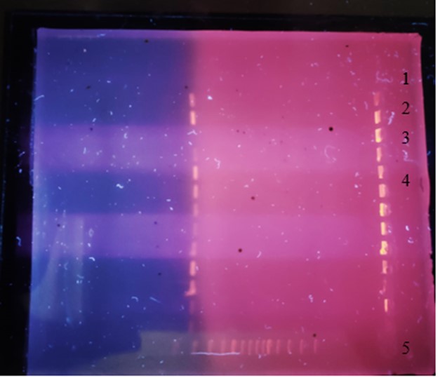
Figure 1. Bands of mecA, 1 Negative control, 2 Positive control, 3 MSSA (mec-A band absent), 4 MRSA (mec-A band present), 5 DNA Ladder
In total 83 patients were included in the study. Their Analysis of 222 samples of Staphylococcus aureus was carried out. In this study using PCR for mec-A gene; MRSA were 32.4% (n=72) and MSSA were 67.56% (n=150), using Cefoxitin; MRSA were 22.5% (n=50) and MSSA were 77.4% (n=172), using Oxacillin; MRSA were 25.6% (n=57) and MSSA were 74.3% (n=165), using Oxacillin Screen agar (OSA); MRSA were 29.2% (n=65) and MSSA were 70.7% (n=157) and by using Oxacillin Agar Dilution; MRSA were 36.9% (n=82) and MSSA were 63.06% (n=140). Keeping in view mec-A gene as gold standard; Cefoxitin disc detected 45 True MRSA (20.2%), Oxacillin disc detected 47 True MRSA (21.17%), OSA detected 59 True MRSA (26.57%) and MIC of Oxacillin detected 66 True MRSA (29.72%) (refer to Table I: True & False Positive & Negative MRSA & MSSA). The results of Phenotypic methods were compared with the results of PCR by using Pearson Chi-square formula.
Table I: True & False Positive & Negative MRSA & MSSA |
||||
Method |
True MRSA |
False MRSA |
True MSSA |
False MSSA |
PCR mecA detection |
72 |
|
150 |
|
Cefoxitin Disc |
45 |
5 |
145 |
27 |
Oxacillin Disc |
47 |
10 |
140 |
25 |
Oxacillin Screen Agar |
59 |
6 |
144 |
13 |
MIC of Oxacillin |
66 |
16 |
134 |
6 |
Table II Sensitivity and Specificity of phenotypic methods |
||
Method |
Sensitivity |
Specificity |
Cefoxitin Disc |
62.5% |
96.6% |
Oxacillin Disc |
65.3% |
93.3% |
Oxacillin Screen Agar |
81.9% |
96% |
MIC of Oxacillin |
91.6% |
89.3% |
The sensitivity and specificity of Cefoxitin Disc Diffusion (CDD) method was 62.5% and 96.6%, Oxacillin Disc Diffusion (ODD) method was 65.3% and 93.3%, Oxacillin Screen Agar (OSA) technique was 81.9% and 96% while that of Oxacillin Agar Diffusion (OAD) was 91.6% and 89.3% respectively (refer to Table II Sensitivity and Specificity of phenotypic methods).
MRSA strains account for a sizable portion of infections in both hospital and community settings due to an increase in the drug resistance patterns.19 MRSA must be accurately identified in order to provide proper treatment and preventive measures. The varied nature of methicillin resistance in Staphylococcus aureus, however, places limitations on the precision and dependability of phenotypic approaches for resistance detection. The mecA gene identification using PCR-based techniques by reference laboratories, are not feasible for routine application in diagnostic settings. Therefore, it is indicated to compare the PCR and phenotypic approaches for the identification of mec-A gene and methicillin resistance. International recommendations like CLSI took many initiatives over the past ten years to standardize and optimize phenotypic approaches for MRSA identification. Since no phenotypic test is 100% reliable for detecting methicillin resistance in Staphylococcus aureus, formation of an algorithm is recommended for accurate identification of MRSA in everyday practice.20
In this study, the evaluation of diagnostic ability of different phenotypic methods like CDD, ODD, OSA and MIC of Oxacillin by agar dilution and their comparison with PCR Assay, showed variable sensitivity and specificity from other international studies. The sensitivity of Cefoxitin disc diffusion is less from most of the studies already conducted that showed sensitivity of almost 100% but specificity is almost similar 100% 21, 100% 22 and 98.3% 23 respectively. The MICs of mec-C resistant organisms cannot be discovered by the tests that target mecA or PBP2a, leading to an increase in percentage of MRSA strains among mec-A negative strains. Strains with mecC are often resistant to cefoxitin and susceptible to oxacillin (CLSI).
In the Oxacillin disc diffusion; results of previous studies 5'24showed sensitivity and specificity of 100% and 73.6% respectively; is quite different from our study showing 65.3% and 93.3% sensitivity and specificity respectively. Similarly the study by 5'22 has higher sensitivity and lower specificity of 84.2% and 66.2% respectively. The reason for decreased sensitivity found in our study might be due to the excessive production of beta-lactamases resulting in phenotypic manifestation of resistance to oxacillin but sensitivity to cefoxitin in mecA gene negative strains, resulting in increase in the number of false positive (MRSA). The Oxacillin Screen Agar results were almost similar to the findings of the studies5,21 which show sensitivity and specificity of 96% and 100% and sensitivity and specificity of 96.3% and 100% respectively. Likewise MIC of Oxacillin by Agar dilution method are close to the findings of 5,25 which shows sensitivity and specificity of 97% and 100% respectively.
The results of our study favored the Oxacillin Screen Agar test for routine MRSA diagnosis and for further confirmation MIC of Oxacillin by Agar Dilution technique. MIC of oxacillin by agar dilution technique is a bit tricky but it detects the maximum number of true positive MRSA strains. Many other methods for MRSA detection are available. Latex agglutination method is a reliable technique having good sensitivity and specificity as shown in the study conducted.26 Epsilometer-strip test (E-Test) is used for determining the MIC of different antibiotics. The results of the studies conducted showed that this test does not provide precise MIC as compared to Broth Microdilution Technique.27 Mannitol-Salt Agar (MSA) detects MRSA by the appearance of yellow-colored colonies on the media after 48 hours, but this technique has a lower sensitivity and specificity as compared to other methods.28
Oxacillin screen agar was demonstrated as the better phenotypic technique for the detection of MRSA in routine practice.
Recommendations: The research can be extended by including these techniques too. Our study serves for the need of extensive research with large sample size from different areas of Pakistan to ascertain the status of different MRSA genes in this part and their comparison with other countries.
- Jayakumar J, Kumar VA, Biswas L, Biswas R. Therapeutic applications of lysostaphin against Staphylococcus aureus. J Appl Microbiol. 2021;131(3):1072–82. https://doi.org/10.1111/jam.14985
- Pal S, Sayana A, Joshi A, Juyal D. Staphylococcus aureus: A predominant cause of surgical site infections in a rural healthcare setup of Uttarakhand. J Fam Med Prim Care [Internet]. 2019 [cited 2022 Aug 22];8(11):3600. https://doi.org/10.4103/jfmpc.jfmpc_521_19
- Tong SYC, Davis JS, Eichenberger E, Holland TL, Fowler VG. Staphylococcus aureus infections: Epidemiology, pathophysiology, clinical manifestations, and management. Clin Microbiol Rev. 2015;28(3):603–61. https://doi.org/10.1128/cmr.00134-14
- Kozajda A, Jeżak K, Kapsa A. Airborne Staphylococcus aureus in different environments—a review. Environ Sci Pollut Res. 2019;26(34):34741–53. https://doi.org/10.1007/s11356-019-06557-1
- Benkő R, Gajdács M, Matuz M, Bodó G, Lázár A, Hajdú E, et al. Prevalence and antibiotic resistance of eskape pathogens isolated in the emergency department of a tertiary care teaching hospital in hungary: A 5-year retrospective survey. Antibiotics [Internet]. 2020 Sep 1 [cited 2021 Jun 30];9(9):1–17. https://doi.org/10.3390/antibiotics9090624
- Tarafder S, Abdullah Yusuf M, Sultana H, Sattar H, Nath Sarker J, Tofael Hossain Bhuiyan M, et al. Comparison of Cefoxitin Disc diffusion test , Oxacillin Disc diffusion test, Oxacillin Screen agar and PCR for mec-A gene for detection of methicillin resistant Staphylococcus aureus.(MRSA). Eur J Pharm Med Res [Internet]. 2019 .
- Huse HK, Miller SA, Chandrasekaran S, Hindler JA, Lawhon SD, Bemis DA, et al. Evaluation of oxacillin and cefoxitin disk diffusion and mic breakpoints established by the clinical and laboratory standards institute for detection of meca-mediated oxacillin resistance in staphylococcus schleiferi. J Clin Microbiol. 2018 Feb 1;56(2). https://doi.org/10.1128/jcm.01653-17
- Harada D, Nakaminami H, Miyajima E, Sugiyama T, Sasai N, Kitamura Y, et al. Change in genotype of methicillin-resistant Staphylococcus aureus (MRSA) affects the antibiogram of hospital-acquired MRSA. J Infect Chemother [Internet]. 2018;24(7):563–9. Available from: https://doi.org/10.1016/j.jiac.2018.03.004
- Dai G, Li Z, Luo F, Lu Y, Chu Z, Zhang J, et al. Simultaneous electrochemical determination of nuc and mecA genes for identification of methicillin-resistant Staphylococcus aureus using N-doped porous carbon and DNA-modified MOF. Microchim Acta [Internet]. 2021;188(2). Available from: https://doi.org/10.1007/s00604-020-04698-6
- Rauf MA, Zafar AM, Amjad AI. Comparison of different phenotypic methods of detection of methicillin resistance in Staphylococcus aureus with the molecular detection of mec-A gene. J Coll Physicians Surg Pakistan. 2008;18(8):529–30.
- Upshaw-Owens M, Bailey CA. Preventing Hospital-Associated Infection: MRSA. MEDSURG Nurs. 2012;21(2):77–81.
- Montazeri EA, Khosravi AD, Jolodar A, Ghaderpanah M, Azarpira S. Identification of methicillin-resistant Staphylococcus aureus (MRSA) strains isolated from burn patients by multiplex PCR. Burns. 2015;41(3):590–4. https://doi.org/10.1016/j.burns.2014.08.018
- Broekema NM, Van Tam T, Monson TA, Marshall SA, Warshauer DM. Comparison of cefoxitin and oxacillin disk diffusion methods for detection of mecA-mediated resistance in staphylococcus aureus in a large-scale study. J Clin Microbiol [Internet]. 2009 [cited 2021 Aug 16];47(1):217–9. https://doi.org/10.1128/jcm.01506-08
- Committee E. EUCAST: Clinical breakpoints and dosing of antibiotics [Internet]. European Committee on Antimicrobial Susceptibility Testing. 2022 [cited 2022 Aug 4]. Available from: https://www.eucast.org/clinical_breakpoints/
- Anand K, Agrawal P, Kumar S, Kapila K. Comparison of cefoxitin disc diffusion test, oxacillin screen agar, and PCR for mecA gene for detection of MRSA. Indian J Med Microbiol [Internet]. 2009;27(1):27–9. https://doi.org/10.1016/S0255-0857(21)01748-5
- Dhungel S, Rijal KR, Yadav B, Dhungel B, Adhikari N, Shrestha UT, et al. Methicillin-Resistant Staphylococcus aureus (MRSA): Prevalence, Antimicrobial Susceptibility Pattern, and Detection of mec A Gene among Cardiac Patients from a Tertiary Care Heart Center in Kathmandu, Nepal. Infect Dis Res Treat. 2021 Jan; 14:117863372110373. https://doi.org/10.1177/11786337211037355
- Lee AYS, Lin M. Clinica Chimica Acta Polymeric IgA paraprotein on agarose gel electrophoresis immunofixation identifies a unique subset of IgA myeloma patients. Clin Chim Acta [Internet]. 2021;512(August 2020):112–6. https://doi.org/10.1016/j.cca.2020.10.031
- Erick MCJ, Miranda G, Sandra D, Argueta E, Wacher NH, Silva M, et al. Evaluation of Different Phenotypic and Genotypic Methods for Detection of Methicillin Resistant Staphylococcus aureus (MRSA). Rev CENIC Ciencias Biológicas [Internet]. 2016;152(3):28. Available from: http://www.ijp.iranpath.org
- Demir T, Coplu N, Esen B. Comparative analysis of phenotypic and genotypic detection of methicillin resistance among Staphylococcus aureus. Indian J Pathol Microbiol [Internet]. 2016 Jul 1 [cited 2022 Sep 27];59(3):314–7. https://doi.org/10.4103/0377-4929.188103
- Kaya EG, Karakoç E, Yaci S, Yücel M. Evaluation of phenotypic and genotypic methods for detection of methicillin resistance in Staphylococcus aureus. African J Microbiol Res [Internet]. 2009;3(12):925–9. Available from: http://www.academicjournals.org/ajmr
- Lohan K, Sangwan J, Mane P, Lathwal S. Prevalence pattern of MRSA from a rural medical college of North India: A cause of concern. J Fam Med Prim Care [Internet]. 2021;10(2):752. https://doi.org/10.4103/jfmpc.jfmpc_1527_20
- Tarafder S, Abdullah Yusuf M, Sultana H, Sattar H, Nath Sarker J, Tofael Hossain Bhuiyan M, et al. Comparison of Cefoxitin Disc diffusion test, Oxacillin Disc diffusion test, Oxacillin Screen agar and PCR for mec-A gene for detection of methicillin resistant Staphylococcus aureus. (MRSA). Eur J Pharm Med Res [Internet]. 2019 [cited 2022 Sep 27];6(10):136–9.
- Nair D, Shashindran N, Kumar A, Vinodh V, Biswas L, Biswas R. Comparison of Phenotypic MRSA Detection Methods with PCR for mecA Gene in the Background of Emergence of Oxacillin-Susceptible MRSA. Microb Drug Resist [Internet]. 2021 Sep 9 [cited 2022 Mar 30];27(9):1190–4. https://www.liebertpub.com/doi/abs/10.1089/mdr.2020.0361
- Farahani A, Mohajeri P, Gholamine B, Rezaei M, Abbasi H. Comparison of different phenotypic and genotypic methods for the detection of methicillin-resistant Staphylococcus aureus. N Am J Med Sci [Internet]. 2013 Nov [cited 2022 Sep 27];5(11):637–40. https://doi.org/10.4103/1947-2714.122305
- Mohanasoundaram KM, Lalitha MK. Comparison of phenotypic versus genotypic methods in the detection of methicillin resistance in Staphylococcus aureus. Indian J Med Res. 2008;127(1):78–84.
- Nair D, Shashindran N, Kumar A, Vinodh V, Biswas L, Biswas R. Comparison of Phenotypic MRSA Detection Methods with PCR for mecA Gene in the Background of Emergence of Oxacillin-Susceptible MRSA. Microb Drug Resist [Internet]. 2021 Sep 9 [cited 2022 Sep 30];27(9):1190–4. https://www.liebertpub.com/doi/10.1089/mdr.2020.0361
- Bushra., Padmaja K, Sudhaharan S, Teja VD. Comparison of Vancomycin MICs by Broth Microdilution Method, E-Test and Vitek 2C among MRSA Isolates in Tertiary Care Centre, Hyderabad, India. Natl J Lab Med. 2022;2019–22.
- Khawaja A, Arshad F, Khan I. Comparison of phenotypic methods with mecA gene-based polymerase chain reaction for Methicillin-resistant Staphylococcus aureus detection. J Pak Med Assoc. 2020;70(2):276–80. https://doi.org/10.5455/jpma.290732

An Official Publication of
Islamabad Medical & Dental College
Vol 13 No (Suppl.)
Address of Correspondence
Nehaj Tariq
Email:
nehajtariq@gmail.com
Authors’ Contribution: 1,2Conception; Literature research; manuscript design and drafting; 3,4 Critical analysis and manuscript review; 5,6Data analysis; Manuscript Editing.
Cite this article. Tariq N, YasmIn S, Butt T, Butt A, Arif S, Bano S. Phenotypic Methods for Detection of Methicillin Resistant Staphylococcus Aureus. J Islamabad Med Dental Coll. 2024; 13i(Suppl.). 499-505. DOI: https://doi.org/10.35787/jimdc.
v13i(Suppl.).1191