Outcome of Operative Vs. Non-Operative Management of Weber B Ankle Fractures
Muhammad Bilal, Adeel Abbass, Muhammad Shakeel Basit, Fahad Aman Khan, Abdul Hannan, Asifa Karamat
Department of Orthopedics, Al Aleem Medical College, Lahore
Objectives: To look at the outcome of Weber B fracture treatment and how various trauma surgeons have been treating them and final outcome with various treatment protocols.
Methodology: Data was collected from medical records after approval of Ethical committee. All ankle fracture presented at two hospitals in Lahore during 2020 to 2023 were evaluated for treatment offered and final outcome was recorded from clinical review and x-rays performed at final visit.
Results: We looked at a total number of 90 Weber B ankle fractures treated at our two institutions during the three years’ time period and conservative vs. operative treatment was offered to the patients as per on call trauma team’s preferred method of treatment. Out of 90 patients, 47 (52%) patients were treated non-operative and 43(48%) patients were operated. 5 patients from non-operative group needed to be operated within first two weeks. All patients went on to achieve full union at the end of one year follow-up.
Conclusion: Our study showed that for non-displaced Weber B ankle fracture, conservative treatment is a suitable alternative to operative management. Risk of treatment failure is very low and patient satisfaction is comparable to operative group.
Keywords: Ankle fracture, ORIF
Ankle fracture is a traumatic injury and very common presentation in orthopedic emergency. It can be high energy trauma as in road traffic accidents or fall from heights. It can also happen after low energy trauma such as twisting injuries in high heels, bad tackle during running and missed steps. Spectrum of injuries ranges from sprains, ligaments tears or bony fracture. Regarding the ankle fractures management is decided by fracture configuration, ligament status and associated injuries. Some injuries are always considered unstable and need surgery such as ankle dislocations, bimalleolar and trimalleolar pattern and syndesmotic displacement.1,2 Stable un-displaced ankle fractures can be treated without surgery with good reported outcomes in literature.3,4 But which ankle fracture will be stable and is there a risk of later instability during the course of treatment. Lot of research has been done
in this area and classifications systems have been proposed. Most common classification followed at our local institute is Weber Classification. Weber proposed this classification based on his experimental work on mechanism of injury, direction of force and its impact on bone components. As per this system, Weber A is minimum displaced fracture of Lateral malleolus and always treated conservatively. Weber C is high energy trauma with unstable pattern and almost always treated with surgical fixation.6,7
Controversy exists regarding management of Weber B type. Un-displaced Weber B can be managed conservatively but there is risk of late displacement and need for surgical management. The stability of trans-syndesmotic Weber B fracture which is also supination external rotation [SER] type fracture remains point for discussion. Lot of research in this area is trying to investigate which trans-syndesmotic Weber B fractures will be potentially unstable. In more than one third of cases of web B injury, the distal tibia fibular syndesmosis is also injured and created ankle joint instability.8 If left untreated this may lead to post traumatic arthritis, chronic pain and worse patent outcomes.9,10 In isolated Weber B injury, deltoid ligament also plays an important role in predicting stability of fracture. Intact deltoid ligament will have better chances of success if treated conservatively. Stress radiographs and clinical examination can be useful in determining the integrity of deltoid ligament. Other factors influence the decision for treatment is degree of displacement, rotation of fracture fragments, patient compliance and patient choice of treatment. Gross displacement, malrotation and poor patient compliance are all indications for surgical fixation to avoid poor outcome in long term. Conservative treatment needs closed follow up and serial radiographs to make sure at fracture position remain within acceptable criteria. If a patient can’t come for follow up then surgical fixation is better choice of treatment.
9,10 In this study, we aim to treat isolated Weber B type fractures with no signs of instability or displacement on x-rays conservatively and report the outcome to compare with national and international standards.11,12 We will look at timing of immobilization, method, follow up and weight bearing status of patients.
We looked at a total number of 90 Weber B ankle fractures treated at our two institutions in Lahore during the three years’ time period and conservative vs. operative treatment was offered to the patients as per on call trauma team’s preferred method of treatment. Aim was to achieve a stable fixation which will allow early weight bearing and rehabilitation.
Table I: Patient Demographics, Treatment Offered, ASA* Grade and Mechanism of Injury |
|
Patient Demographics |
|
Male |
64 |
Female |
26 |
Age Range |
19-72 |
Average Age |
44 |
Treatment Offered |
|
Conservative |
47 – 5 converted patients=42 |
Operative |
43 + 5 addition=48 |
Converted from conservative |
5 |
Total operative cases |
48 |
ASA Grade |
|
ASA Grade I |
28 |
ASA Grade II |
17 |
ASA Grade III |
3 |
Mechanism of Injury |
|
Trip and fall |
30 |
Fall from height |
5 |
Bike accident |
40 |
Trivial trauma |
10 |
Other |
5 |
Minimum planned follow up was one year since time of surgery for all patients. Patient ward notes and outpatient follow ups were carefully recorded for identification of any signs of instability, change in fracture configuration, non-union, chronic pain, and post-traumatic arthritis. Weber Ankle Fracture classification system was used to define the fracture type and documented. 11,12
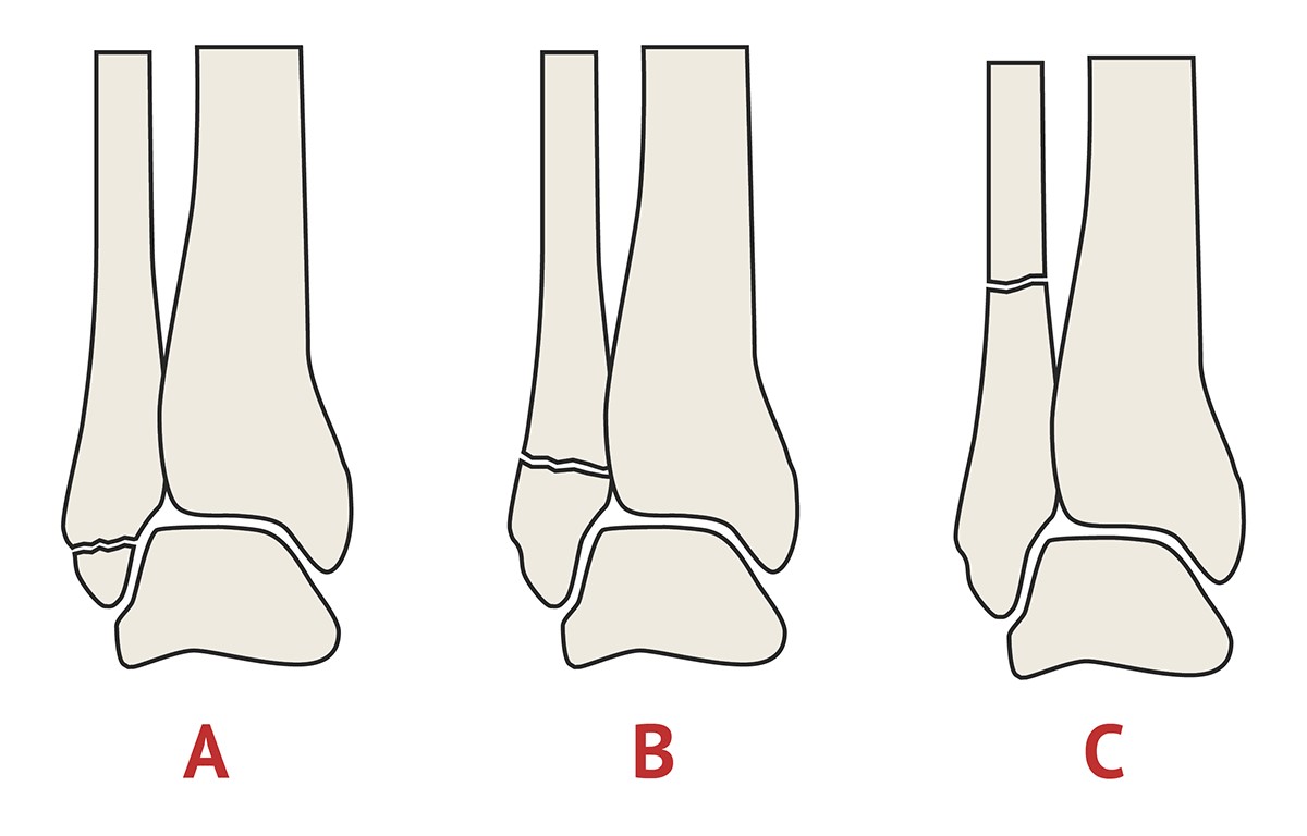
Figure 1. Weber Type A and B and C as per Weber Ankle Fracture Classification
At last decided follow up, fracture healing and overall outcome was documented as excellent (18 points), good (15–17points), fair (13–14 points) or poor (< 13 points) in terms to the modified Merle d’Aubigné-Postel score and radiological healing was scored as excellent, good, fair, or poor based on Matta score.13,15
We treated total number of 90 patients with diagnosis of Weber B ankle fracture over the period of three years in two hospitals. Out of 90 patients, 47 (52%) patients were treated non-operative and 43 (48%) patients were operated. 5 patients from non-operative group needed to be operated within first two weeks. All patients went on to achieve full union at the end of one year follow-up.
Significant finding was noted that at the final clinical review there was no difference in patient satisfaction and fracture healing in both groups. Surgery was performed within two weeks of initial presentation and patient was given time for surgery on the trauma lists. While conservative group was discharged within 48 hours and then followed up at one week, two week and 6 weeks intervals. Weight bearing x-rays were performed at one week and if fracture stable, conservative treatment was continued with completion of POP cast. If at first or subsequent follow up any fracture displacement was noted, then surgical treatment was offered to the patients.
All patients were followed up for 12 months. Two surgical site infections was reported which was successfully treated with antibiotics. Average surgical fixation time was 45 minutes. Blood loss was minimum. We used standard Venous Thromboembolism protocol for our post-operative patients and none of the patient developed deep vein thrombosis (DVT) or Pulmonary embolism (PE). At final follow up no fixation failure was noted although minor loss of reduction was noted on serial radiographs. This was evaluated according to the criteria proposed by Matta.13 All fractures were united at an average of 3 ± 2 months (range, 4–7 months). Although conservative group was in protected weight bearing in a plaster cast or ankle boot for 2 extra weeks compared to operative group. Among all patients, the clinical outcomes were recorded. In operative group, patients reported as excellent in 31 patients (31/48, 65%), good in 9 (9/48, 19%), fair in 3 patients (3/48, 7%) and poor in 4 (4/48. 9%). Poor outcome was mainly due to infection in surgical patients and one patient developed complex regional pain syndrome.15, 16
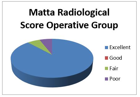
Figure 2: Radiological score of Operative Group (Matta Scoring System)
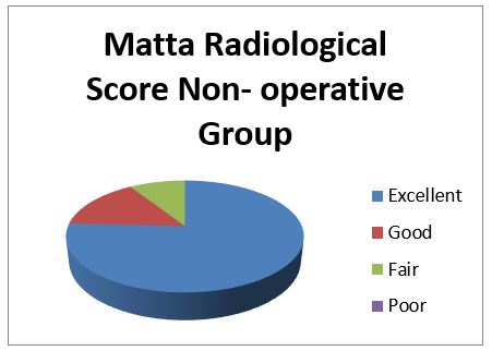
Figure 3: Radiological score of Non-Operative Group (Matta Scoring System)
Table II. Operative vs Non-Operative Outcome of Fracture |
||
Fracture Outcome |
Operative |
Non-Operative |
Total patients |
43 |
47 |
Anatomical Reduction Achieved |
40 |
30 |
Displaced Reduction |
3 |
12 |
Poor Reduction |
0 |
5 converted to surgery |
Regarding the non-operative group patients reported as excellent in 34 patients (34/42, 80%), good in 6 (6/42, 13%) and fair in 2 patients (2/42, 7%). None of the patients reported poor outcome but five patients were converted from no-operative group to operative group due to displacement of fracture alignment. The average modified Merle d’Aubigné-Postel score was 15 ± 2.0 points (range, 10–18 points).
Table III. Comparison of fracture healing and patient reported outcome of 2 groups |
|||
Outcome measure |
Operative group |
Non-operative group |
P Value |
Average Age |
50.7 (41.1–60.5) |
45 (25.7–58.2) |
0.3 |
Average BMI |
28(24-30) |
26(23-28) |
0.09 |
No of Complications |
2 (Infection) |
5 (converted to surgery) |
0.7 |
Average number of Follow ups |
6 |
5 |
0.5 |
Average Duration of Immobilisation |
4weeks |
6weeks |
0.3 |
Average number of x-rays |
3 |
5 |
0.6 |
(P-value was calculated using chi-square test. P-value is considered significant <0.05.) The radiological healing was assessed according to the Matta score [22] and were recorded for both groups. For operative group, it was Excellent 45 patients (45/48, 94%) and fair in 3 patients (3/48, 7%). No poor healing was recorded in operative group. For non-operative group, five patients were converted to surgery due to loss of reduction. In the remaining population healing and fracture position was recorded as excellent in 25 patients (25/42, 60%), good in 5 (5/42, 12%) and fair in 12 patients (12/42, 28%). All patients achieved complete union and no poor outcome was recorded. Figure 2 and 3.
Ankle fracture following trauma is a very common injury and all orthopedic surgeons are familiar with several algorithms to treat these injuries. After latest trend of minimal surgical interventions ankle fracture are also under the spotlight to achieve best outcomes with minimum possible interventions. Regarding fracture pattern, there is agreement between the surgeons that isolated lateral malleolar fractures with intact medial column are stable injuries and can be treated conservatively in plasters or functional boots with regular monitoring. Our study was based on this objective and we successfully treated 95% patients with conservative treatment.16 Several studies have been published which recommend use of plasters or functional bracing for un-displaced type B ankle injuries such as Observational Study of Surgery for type-B Ankle Fracture Treatment [CROSSBAT],13 British Orthopaedic Association Standards for Trauma [BOAST] guidelines 2016.14 Clinical concern regarding fracture displacement or missed injuries remain. However, some understandable concern remains among trauma surgeons regarding identification of stable ankle fracture. As such fractures may still displace if patients are allowed to weight bear in a brace.17 As in our study population five patients went on from stable fracture configuration to displaced and were operated on within two weeks. Therefore, a variety of clinical signs and observations are used to look out for stable fracture which can be successfully treated without surgery. Such as high energy trauma, medial malleolar pain, bruising, positive stress test and displacement on weight bearing radiographs. Medial tenderness could be a mild degree of deltoid sprain and can still be managed conservatively.17 Stress x-rays are difficult to perform in acute pain and can be misleading if not properly done.18 On the other hand, with proper training of radiographers Weight bearing x-rays can be more useful to detect unstable injuries. In his series of 57 patients, Weber et al. conducted a study on 57 patients and described the weight bearing radiographs as easy to obtain and reproducible check for instability.19,20 conducted weight bearing x-rays in all patients before deciding for treatment choice. And these x-rays were helpful in decision making process.17 Treatment of stable ankle fracture in functional bracing 20 is years old. Stuart et al reported good results in these patients using pneumatic stirrups.16 Another study also documented patient comfort and function in bracing and conservative treatment of stable fractures.18 Our study did not show any significant statistical difference in the fracture union or patient reported outcome score. All our patients achieved complete union at final follow up. However, the surgical group was allowed to start weight bearing 2 weeks earlier than conservative group. But due to surgery, clinical appointments were more in operative patients.
Limitations of our study are retrospective data, no randomization and small number of patients. A randomized control trial in multiple hospitals will give more insight into conservative management of stable ankle fractures.14,18
Ankle fractures are very common and treated in multiple ways. We aimed to look at the outcome of conservative treatment of stable type B Weber fracture. Our study shows that non-operative treatment produces satisfactory outcome in this injury and can be continued in trauma setting.
- Jennison T, Brinsden M. Fracture admission trends in England over a ten-year period. Ann R Coll Surg Engl 2019;101(3):208–14. https://doi.org/10.1308/rcsann.2019.0002
- Javed OA, Javed QA, Ukoumunne OC, Di Mascio L. Surgical versus conservative management of ankle fractures in adults: a systematic review and meta-analysis. Foot Ankle Surg 2019;26(7):723–35. https://doi.org/10.1016/j.fas.2019.09.008
- Larsen P, Rathleff MS, Elsoe R. Surgical versus conservative treatment for ankle fractures in adults—a systematic review and meta-analysis of the benefits and harms. Foot Ankle Surg 2019;25(4):409–17. https://doi.org/10.1016/j.fas.2018.02.009
- Kortekangas T, Haapasalo H, Flinkkil¨a T, Ohtonen P, Nortunen S, Laine H-J, et al. Three week versus six week immobilisation for stable Weber B type ankle fractures: randomised, multicentre, non-inferiority clinical trial. BMJ 2019;364: k5432.https://doi.org/10.1136/bmj.k5432
- Elgayar L, Arnall F, Barrie J. A systematic review investigating the effectiveness of surgical versus conservative management of unstable ankle fractures in adults. J Foot Ankle Surg 2019;58(5):933–7. https://doi.org/10.1053/j.jfas.2018.12.017
- Lampridis V, Gougoulias N, Sakellariou A. Stability in ankle fractures: diagnosis and treatment. EFORT Open Rev 2018;3(5):294–303. https://doi.org/10.1302/2058-5241.3.170057
- Bonness EK, Siebler JC, Reed LK, Lyden ER, Mormino MA. Immediate weight-bearing protocol for the determination of ankle stability in patients with isolated distal fibular fractures. J Orthop Trauma 2018;32(10):534–7.https://doi.org/10.1097/bot.0000000000001268
- Schnetzke M, Vetter SY, Beisemann N, Swartman B, Grützner PA and Franke J: Management of syndesmotic injuries: What is the evidence? World J Orthop7(11): 718-725, 2016.https://doi.org/10.5312/wjo.v7.i11.718
- Kortekangas T, Flinkkila T, Niinimaki J, Lepojarvi S, Ohtonen P, Savola O and Pakarinen H: Effect of syndesmosis injury in SER IV (Weber B)-type ankle fractures on function and incidence of osteoarthritis. Foot Ankle Int 36(2): 180-187, 2015.https://doi.org/10.1177/1071100714551788
- Veen EJ and Zuurmond RG: Mid-term results of ankle fractures with and without syndesmotic rupture. Foot Ankle Surg 21(1): 30-36, 2015.https://doi.org/10.1016/j.fas.2014.09.001
- Manjoo A, Sanders DW, Tieszer C and MacLeod MD: Functional ad radiographic results of patients with
- syndesmotic screw fixation: implications for screw removal. J Orthop Trauma 24(1): 2-6, 2010.https://doi.org/10.1097/bot.0b013e3181a9f7a5
- Jung HG, Kim JI, Park JY, Park JT, Eom JS and Lee DO: Is hardware removal recommended after ankle fracture repair? Biomed Res Int 2016: 5250672, 2016.https://doi.org/10.1155/2016/5250672
- Inam M, Palh MB, Raza T,Alam F, Ahmed I, Abidi SA et al. (2024). Acetabular Fracture; its Management and Early Functional Outcome. Pakistan Journal of Medical & Health Sciences, 18(01), 51. https://doi.org/10.53350/pjmhs0202418151
- Stuart PR, Brumby C, Smith SR. Comparative study of functional bracing and plaster cast treatment of stable lateral malleolar fractures. Injury 2019;20(6): 323–6. https://doi.org/10.1016/0020-1383(89)90003-x
- Murphy, E.A., Ehrhardt, B., Gregson, C.L. et al. Machine learning outperforms clinical experts in classification of hip fractures. Sci Rep 12, 2058 (2022). https://doi.org/10.1038/s41598-022-06018-9
- Van den Berg C, Haak T, Weil NL, Hoogendoorn JM. Functional bracing treatment for stable type B ankle fractures. Injury 2018;49(8):1607–11. https://doi.org/10.1016/j.injury.2018.06.009
- Kearney RS, McKeown R, Stevens S, Parsons N, Parsons H, Wells P, et al. Cast versus functional brace in the rehabilitation of patients treated for an ankle fracture: protocol for the UK study of ankle injury rehabilitation (AIR) multicentrerandomised trial. BMJ Open 2018;8(12):e027242. https://doi.org/10.1136/bmjopen-2018-027242
- Gregersen MG. Weight-bearing radiographs to evaluate stability in ankles with isolated weber type B fractures. https://doi.org/10.1177/10711007211027286.

An Official Publication of
Islamabad Medical & Dental College
Vol 13 No (Suppl.)
Address of Correspondence
Muhammad Bilal
Email:
drbilalfrcs@yahoo.com
Authors’ Contribution: 1,2Conception; Literature research; manuscript design and drafting; 3,4 Critical analysis and manuscript review; 3,4Data analysis; Manuscript Editing.
Cite this article. Bilal M, Abbas A, Basit MS, Khan FA, Hannan A, Karamat A. Outcome of Operative Vs. Non-Operative Management of Weber B Ankle Fractures. J Islamabad Med Dental Coll. 2024; 13i(Suppl.). 554-559
DOI: https://doi.org/10.35787/jimdc.
v13i(Suppl.).1197