Trends of Antimicrobial Sensitivity among Common Isolates in a Tertiary Care Hospital, Rawalpindi
Uzma Mussarat1, Shazia Taj2, Shahina Yasmin1, Aleena Butt1, Nehaj Tariq3, Tahira Inam1
1Riphah International University, Islamabad, 2National University of Sciences and Technology, Islamabad, 3WATIM Medical & Dental College, Rawalpindi
Objective: To evaluate the resistance patterns of frequently found pathogens to different antimicrobial agents, aiding physicians in choosing appropriate treatment strategies for patients within our locality.
Methodology: This retrospective observational study took place at Pakistan Railway hospital (PRH) Rawalpindi; from January 2021 to December 2021. Data were obtained from the institute's database, the Hospital Information Management System (HIMS). Pathogens were identified in the laboratory through Gram staining and biochemical tests, and their antibiotic sensitivity profiles using disk diffusion method assessed following protocols established by the Clinical & Laboratory Standards Institute. Statistical analysis was conducted using SPSS version 24, and data entry was carried out accordingly.
Results: Overall, 1128 positive cultures were reported in study period including urine, pus, HVS, sputum cultures, stool, blood, catheter tip, and others (tissue fluid, wound, and cannula tip etc.). They comprised 32.8 %, 30.9 %, 16.6%, 5.05 %, 3.81%, 3.72 %, 1.15%, 0.70 % and 0.08% of the other specimens respectively. Escherichia coli was the most prevalent gram-negative species 391(34.66%) followed by Klebsiella pneumoniae 247 (21.89 %). Among the isolates, Escherichia coli revealed 43% resistance against Cefotaxime (one of the third-generation Cephalosporin) and Klebsiella pneumoniae exhibited 49% resistance to Cefotaxime. Moreover increased resistance was also noted against Penicillin’s and Quinolones by both E.Coli and Klebsiella species.
Conclusion: Moderate to reduced susceptibility to frequently utilized antimicrobials has been noted across diverse gram-positive and gram-negative species, apart from Penicillin. To address the escalating resistance to Carbapenems and Cephalosporin, it is essential to establish and enforce rigorous infection control measures.
Keywords: antimicrobial resistance, microbial pathogens, patient safety
The emergence of antibiotics in 1928 when Alexander Fleming invented penicillin, is often acclaimed as one of the peak significant medical advancements of the past era.1 A solitary dose of Penicillin had the potential to eliminate life-threatening infections. Nevertheless, the pervasive issue of antimicrobial resistance looms large, posing a serious threat to nullify this historic achievement and potentially ushering in a post-antibiotic era.2 Emerging antimicrobial resistance (AMR) is a major threat in today’s world in both developed and developing countries. World Health Organization (WHO) has declared AMR among top ten fears to global health.3 This evolving hazard is becoming a main dynamic milestone for long duration hospital stay, augmented problems related with disease, significant economic burden and an overall venomous influence on health.2 Regular analysis of infection patterns and their antibiogram in resource-poor settings specially is crucial for pragmatic management. Frequent use of antimicrobials over the past 20 years has led to changed susceptibility pattern of frequently found pathogens.4 Developing countries including Pakistan are the major consumers of antibiotics that contribute to deprived observance to infection control trials, injudicious consumption of antibiotics, and inadequate antimicrobial formulations. A strong surveillance system is obligatory to address the global concern of AMR. Preferably, antibiotics should be prescribed according to AMR surveillance treatment guidelines but conflicting to this detail patients with bacterial infections in evolving countries are generally controlled analytically. This emphasizes the requirement for timely and regular informs of the continually switching drug resistance patterns5. Avoidance of record related to accelerated resistance of frequently used antimicrobials can lead to increased morbidity and mortality.5 In 2011, the World Health Organization (WHO) designated antimicrobial resistance (AMR) as the focal idea for World Health Day. The campaign's mantra, "Combat Drug Resistance – No action today, no cure tomorrow," emphasized the urgency of addressing this issue. To support this initiative, six- policy points were instituted as a central component for World Health Day.6
Present study is directed to comment on existing microbial pathogens and their antimicrobial susceptibility patterns among inpatients as well as outpatients to guide antimicrobial stewardship as well as to implement patient safety programs for improved patients’ care. The study will help the policymakers, Physicians, and the overall population on the magnitude of AMR in this setting especially in developing countries where monitoring and controlling AMR is predominantly challenging.
This retrospective observational study was conducted at Microbiology section of Pathology department at Pakistan Railway Hospital (PRH) from January 2021 to December 2021. The study was selected by the Review Committee of Islamic International Dental College (Ref # Riphah/ IIDC/ IRC/2023 /003/002). A total of 1128 cultures with growth of microbial pathogens were included in the study. Samples with positive cultures from both indoor and outdoor patients (Urine, pus, HVS, sputum cultures, stool, Catheter tip, blood, and others (tissue fluid, wound, and cannula tip etc.), irrespective of age and gender were included in the study were included in inclusion criteria. Samples without culture growth were excluded. The isolates were identified with the aid of standard microbiological specimen detection protocols. Culture of bacterial samples, Gram staining and succeeding identification of bacterial isolates were accomplished by trained microbiologists. Routine biochemical testing performed to confirm the bacterial pathogens. Kirby-Bauer Disc Diffusion method was preferred for Antibiotic Susceptibility Testing. A suspension was prepared from growth on a solid media plate by adding bacterial colonies into sterile distilled water. The bacterial suspension was made attaining turbidity level of 0.5 McFarland turbidity standard and inoculated on Muller Hinton agar plates. Subsequent step was placement of antibiotic disks on agar plate with a minimum separation of 20 mm and incubated for 18–24 hours at 37 °C. The zones of inhibition were calculated in millimeters and the isolates were categorized as “resistant,” “intermediate,” or “sensitive” according to the guidelines provided by the Clinical and Laboratory Standards Institute (CLSI)7,8. An Antibiogram was carried out by using the following groups of antibiotics disks: Ampicillin (AMP 10 μg), Amoxicillin/Clavulanic acid (AMC 20/10 μg), Amikacin (AK 30 μg), Azithromycin (15 μg), Aztreonam (30 μg), Cefotaxime (CTX 30 μg), Ceftazidime (CAZ 30 μg), Ceftriaxone (CRO 30 μg), Ciprofloxacin (CIP 5 ug), Cefuroxime (30 μg), Trimethoprim/Sulfamethoxazole (1.25/23.75 μg), Cloxacillin (CLOX 5 μg), Vancomycin (VAN 30 μg), Erythromycin (E 10 μg), Imipenem (IPM 10 μg), Penicillin (10 μg), Ofloxacin (5 μg), Gentamicin (CN 10 μg), Tetracycline (TET, 30 μg), Fosfomycin (FOX 50 μg), and Nitrofurantoin (F 300 μg), Clindamycin (DA, 15 μg), Meropenem (MEM 10 μg), Chloramphenicol (C, 30 μg)
Data were entered by the Statistical Package for the social sciences (SPSS) version 24 for evaluation. For qualitative variables (sexual category of patient, category of samples, organisms identified, ward, and antimicrobial susceptibility.) simple descriptive statistics (frequencies, percentages) were calculated. Descriptive numerical (continuous) variables of age (years) were calculated in terms of Mean ± SD (standard deviation).
Overall, 1128 positive cultures were obtained from specimens being processed in Microbiology Lab over the study period. Cultures of urine, pus, HVS, sputum, blood, catheter tip, and others (tissue fluid, wound, and cannula tip etc.) comprised of 370 (32.8 %), 349(30.9 %), 188(16.6%), 57(5.05 %), 43(3.81%), 42(3.72 %), 13(1.15%), 8(0.70 %) and 1(0.08%) of the other specimens respectively. Distribution of isolated pathogens among positive cultures is shown in Table I, As shown in the table Escherichia coli was the most frequently found pathogen [391 (34.66%)]. The second most common bacteria were Klebsiella pneumoniae counted as 247 (21.9%) of total culture positive specimens. Mean age (years) of the patients was 44.22 years with Std. Deviation ± 19.682. Most of the subjects was between 53 to 55 years of ages. The age distribution of different age groups is between 1 to 89 yrs. as shown in Figure 1.
Table 1: Frequency of individual Gram positive and Gram-negative isolates (n=1128) |
||
No |
Pathogen |
Number (%) |
1 |
Escherichia coli |
391 (34.66) |
2 |
Klebsiella pneumoniae |
247 (21.89) |
3 |
Staphylococcus aureus |
201 (17.81) |
4 |
Acinetobacter baumannii |
102 (9.04) |
5 |
Methicillin resistant staphylococcus aureus (MRSA) |
61 (5.40) |
6 |
Pseudomonas aeruginosa |
54 (4.78) |
7 |
Enterococcus faecalis |
35 (3.10) |
8 |
Providencia stuartii |
15 (1.32) |
9 |
Proteus vulgaris |
8 (0.70) |
10 |
Shigella flexneri (3) & S. soneii (4) |
7 (0.62) |
11 |
Salmonella typhi (2) & S. Paratyphi (2) |
4 (0.35) |
12 |
Citrobacter |
3 (0.26) |
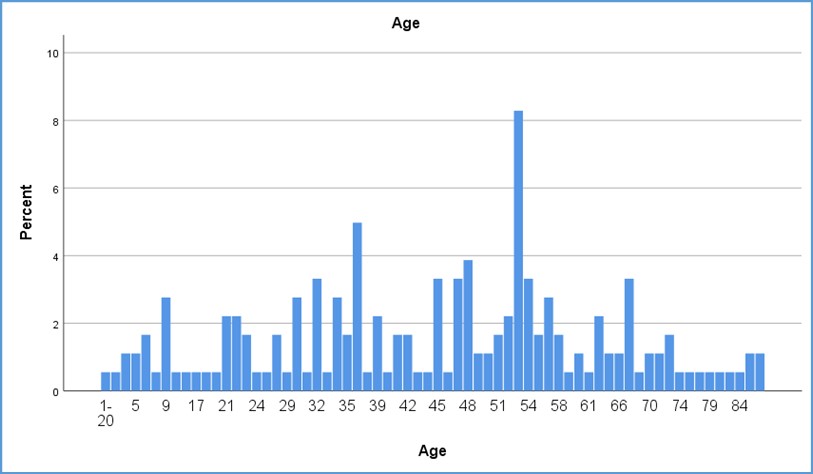
Figure 1: Age distribution of Study Participants
Resistance profile of Gram positive & negative isolates in culture positive specimen is shown in Table II and their comparison of susceptibility is revealed via stacked columns in Figure 2 & 3
Table II: Antibiotic resistance profile of Gram positive & negative isolates in culture positive specimen |
||||||||
No |
Antibiotics |
Isolates |
||||||
Stap. Aureus |
MRSA |
Enterococcus faecalis |
E. coli |
K. pneumoniae |
A. baumannii |
P. aeruginosa |
||
R (%) |
R (%) |
R (%) |
R (%) |
R (%) |
R (%) |
R (%) |
||
1 |
AMP |
180 (97.8) |
46 (75.4) |
10(29) |
226 (58) |
152 (62) |
28(27) |
4(07) |
2 |
AMC |
35 (19.0) |
18(30) |
3(9) |
131(34) |
95(38) |
6(06) |
4(07) |
3 |
AUG |
2 (1.0) |
1(2) |
0(0) |
12(03) |
3(01) |
0(0) |
5(09) |
4 |
AK |
7(3.8) |
2(3) |
1(03) |
40(10) |
33(13) |
8(08) |
0(0) |
5 |
Azactam |
23(12.5) |
17(27.9) |
6(17) |
30(08) |
17(07) |
8(08) |
7(13) |
6 |
AZT |
3(1.6) |
0(0) |
2(06) |
19(05) |
29(12) |
2(02) |
15(28) |
7 |
CTX |
30(16.3) |
13(21.3) |
2(06) |
168(43) |
120(49) |
16(16) |
2(04) |
8 |
CIP |
39(21.1) |
27(44) |
8(23) |
121(31) |
57(23) |
17(17) |
16(30) |
9 |
COT/SXT |
24(13.04) |
18(30) |
6(17) |
72(18) |
60(24) |
8(08) |
7(13) |
10 |
CN |
24(13.04) |
34(56) |
1(03) |
51(13) |
35(14) |
13(13) |
12(22) |
11 |
CLOX |
13(7.0) |
22(36) |
2(06) |
17(04) |
13(05) |
0(0) |
8(15) |
12 |
C |
0(0) |
0(0) |
0(0) |
35(09) |
33(13) |
2(02) |
2(04) |
13 |
Cro |
1(0.5) |
2(3) |
0(0) |
34(09) |
12(05) |
2(02) |
1(02) |
14 |
CE |
0(0) |
3(5) |
0(0) |
7(02) |
5(02) |
1(01) |
1(02) |
15 |
CAZ |
9(4.8) |
7(11) |
8(23) |
11(03) |
3(01) |
3(03) |
9(17%) |
16 |
DOX |
19(10.3) |
10(16) |
8(23) |
28(07) |
17(07) |
3(03) |
5(09%) |
17 |
E |
14(7.6) |
8(13) |
1(03) |
18(05) |
16(06) |
3(03) |
2(04%) |
18 |
FOS |
3(1.6) |
3(05) |
3(09) |
30(08) |
19(08) |
3(03) |
0(0%) |
19 |
F/NIT |
5(2.71) |
0(0) |
1(03) |
22(06) |
26(11) |
3(03) |
1(02%) |
20 |
IPM |
2(1.0) |
1(2) |
1(03) |
4(01) |
1(00) |
6(06) |
6(11%) |
21 |
LZD |
2(1.0) |
1(2) |
0(0) |
Not Applied |
|||
22 |
MEM |
3(1.6) |
1(2) |
0(0) |
21(05) |
9(04) |
6(06) |
3(06) |
23 |
SCF |
8(4.3) |
3(5) |
3(09) |
36(09) |
15 |
7(07) |
4(07) |
24 |
TET |
9(4.8) |
7(11) |
8(23) |
2(01) |
2(01) |
0(0) |
1(02) |
25 |
VA |
38(20.6) |
15(25) |
11(31) |
Not Applied |
|||
26 |
TZP |
6(3.2) |
1(2) |
0(0) |
17(04) |
30(12) |
3(03) |
7(13) |
AMC (Ampicillin/Clavulanic acid), AK(Amikacin), CIP(Ciprofloxacin), LEV(Levofloxacin), GEN (Gentamicin CRO(Ceftriaxone), FEP(Cefepime), TPZ(Tazobactam/Piperacillin), COL (Colistin), NIT(Nitrofurantoin), MIN(Minocycline), IPM(Imipenem), MEM(Meropenem), DOX(Doxycycline), SXT (trimethoprim/sulfamethoxazole), CAP(Chloramphenicol), CAZ (Ceftazidime), TGC (tigecycline)
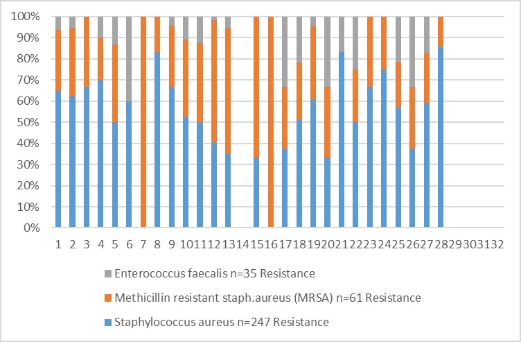
Figure 2: Antimicrobial sensitivity of Gram-positive pathogens
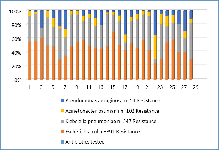
Figure 3: Antimicrobial sensitivity of Gram-negative pathogens
The susceptibility patterns of pathogens have evolved over time, highlighting the necessity for regular monitoring to reduce therapeutic failures and intensify efforts to combat the escalating prevalence of antibiotic resistance. Antimicrobial resistance (AMR) has surfaced as a significant public health issue in both emerging and progressed nations, prompting the World Health Organization (WHO) to advocate for continuous surveillance of AMR.9 Regardless of this insistent demand, a minority of studies till date have conveyed resistance inclinations in different areas of Pakistan.10 Data presented in this report provides a detailed scrutiny of antimicrobial resistance. In our study the highest percentage of Gram-positive and Gram-negative isolates were found and majority of patients were up to 55 years of age group. Almost similar results have been reported by Asad Ullah et al. Their highest percentage was 50 to 59 years of age group.11 Our results were contrary with Siddiqui et al.12 These percentages can fluctuate based on various factors including the study population, patient immunity, specimen collection method, and duration of the study period. In the current study, it was observed that urinary tract infection (UTI) poses a notable challenge, with E. coli being the most frequently reported pathogen exhibiting high resistance to first-line antibiotics. Similar findings have been reported in multiple studies conducted in Pakistan, Bangladesh, and Africa, indicating a widespread issue of inappropriate antibiotic usage in developing nations.10 Our examination of antimicrobial resistance (AMR) among bacterial isolates unveiled drug resistance across both gram-negative and gram-positive organisms, aligning with findings from other studies.13,14 There were also superior rates of co-resistance to universally used antibiotics like Quinolones, and Aminoglycosides and a few isolates showed resistance to Imipenem. Present study discloses that majority (97%) of S. aureus isolates was resistant to Ampicillin, and 20.6% were also resilient to Vancomycin. These findings are consistent with a study that reveal majority (82%) of S. aureus was resistant to penicillin’s and 6% resistant to Vancomycin.13 In present data a significant decrease in antimicrobial resistance rates is revealed to Augmentin and Cephalosporin except Cefotaxime, albeit much lower than those observed for amoxicillin and penicillin, these facts are contrary as seen by other study conducted at Zimbabwe.15 This rise may be attributed to an elevated usage of these drugs by clinicians who aim to circumvent the highly resistant amoxicillin and penicillin. In our study, E. coli emerged as the most frequently isolated pathogen in urine samples (34.66%), followed by Klebsiella spp. (21.89%). This discovery mirrors findings from other studies, where E. coli tends to be the primary uropathogen, followed by Klebsiella species.16,17 In our investigation, E. coli exhibited the highest resistance among bacterial pathogens to first-line antibiotics in the majority of studies. E. coli displayed a high resistance to penicillin (Ampicillin and Amoxicillin), Ciprofloxacin and Cefotaxime while maintaining susceptibility to Colistin, Cefoperazone -Sulbactam, Imipenem, and Meropenem, Tetracycline and Vancomycin. These findings closely align with previous studies conducted in Bangladesh and Africa, indicating a common trend of antibiotic misuse in developing countries.18 Our study revealed elevated median resistance to Tetracycline and Levofloxacin compared to study from Bangladesh. This variance could potentially be attributed to differences in antimicrobial resistance testing methods.18 Our research highlights extensive resistance among both gram-negative and gram-positive bacterial pathogens responsible for common infections like urinary and respiratory tract infections, irrespective of whether they are community-acquired or hospital-acquired. This aligns with findings from other studies in the United States, which indicate that within hospital settings, gram-negative pathogens such as Klebsiella pneumoniae, Pseudomonas aeruginosa, Escherichia coli, and Acinetobacter baumannii demonstrate a high prevalence and strong association with antimicrobial resistance, as evidenced by recent studies.19,20 A study conducted in a tertiary care hospital Wah Cantt by Lubna et al. demonstrated high resistant pattern of Acinetobacter baumannii (89.9%, 78.9%) and Pseudomonas aeruginosa (80%, 60%) to gentamicin and amikacin respectively. While moderate resistance was observed against Klebsiella pneumoniae (53.3%, 46.6%) and Enterobacter spp. (37.5%) for both gentamicin and amikacin.21 These findings are not consistent with our study which reveal only MRSA presented 56% presented to these mentioned drugs. In the past, Carbapenems were considered effective against multidrug-resistant (MDR) Acinetobacter baumannii. However, with the emergence of pan-resistant strains of Acinetobacter baumannii, treating infections caused by this pathogen has become even more challenging. Nevertheless, the level of drug resistance varies significantly across different regions of the world. In our study Carbapenem resistance against Acinetobacter baumannii is very low as compared to the data shared by other countries, the reason could be limited use of this group of medicine in study population. These results are contrast to a study conducted in Rwanda, which found a notable increase in direct resistance to Imipenem among Klebsiella, E. coli, Pseudomonas, and Acinetobacter isolates.22 Hussain et al's studies documented a 70% resistance rate of MRSA to Levofloxacin, while Mekonnen et al.'s reported increased resistance rate to Quinolones in Ethiopia.23,24 In our study, there was a high resistance rate to Ampicillin (75.4%). These frequencies may differ due to influences such as age, immune status, site, and seriousness of infection, as well as geographical variances.
Antimicrobial resistance (AMR) poses a significant global health risk. Effective management of multidrug-resistant (MDR) Gram-negative pathogens hinges upon understanding the spread of AMR and monitoring trends in hospital-acquired infections. Our study's antibiogram indicates an increased resistance to penicillin, cephalosporins, and gentamicin, with emerging resistance to other commonly prescribed medications. However, these may be regional disparities to similar studies should be conducted periodically in different regions in neighboring hospitals. These regional disparities in resistance rates underscore the role of antimicrobial selection and irrational usage in fostering early resistance to cost-effective drugs effective against minor infections. The significance and indispensability of antibiotics in treating infectious diseases cannot be overstated; they are pivotal and should never be viewed merely as commodities.
Future Recommendations
MDR presents a distinctive challenge, emphasizing the need for ongoing monitoring of disease control procedures and regular assessment of antimicrobial susceptibility profiles within our healthcare facilities. There should be a collaborative effort between microbiologists and clinical practitioners to introduce updated and appropriate antimicrobial agents aligned with the evolving trends in antimicrobial resistance (AMR) and locally established antibiograms. To mitigate the risk of early resistance emergence against existing drugs, active surveillance of antimicrobial susceptibility patterns among local isolates should be conducted routinely. Additionally, it's crucial to develop effective empirical therapies to ensure judicious use of antimicrobials. Encouraging the implementation of institutional drug policies, antibiotic stewardship programs, and adherence to stringent infection control practices are also imperative steps in addressing this challenge.
- Mulani MS, Kamble EE, Kumkar SN, Tawre MS, Pardesi KR. Emerging strategies to combat ESKAPE pathogens in the era of antimicrobial resistance: a review. Frontiers in microbiology. 2019;10:539. https://doi.org/10.3389/fmicb.2019.00539.
- Karlowsky JA, Hoban DJ, Hackel MA, Lob SH, Sahm DF. Resistance among Gram-negative ESKAPE pathogens isolated from hospitalized patients with intra-abdominal and urinary tract infections in Latin American countries: SMART 2013-2015. Brazilian Journal of Infectious Diseases. 2017;21:343-8. https://doi.org/10.1016/j.bjid.2017.03.006. Epub 2017 Apr 8.
- Sheikh S, Vishwas G, Aggarwal M, Bhattacharya S, Kumari P, Parashar L, et al. Antibiotic point prevalence survey at a tertiary healthcare hospital in India: Identifying strategies to improve the antibiotic stewardship program immediately after a COVID-19 wave. Infection Prevention in Practice. 2022;4(4):100253. https://doi.org/10.1016/j.infpip.2022.100253
- Kumburu HH, Sonda T, Mmbaga BT, Alifrangis M, Lund O, Kibiki G, et al. Patterns of infections, aetiological agents and antimicrobial resistance at a tertiary care hospital in northern Tanzania. Tropical Medicine & International Health. 2017;22(4):454-64. https://doi.org/ 10.1111/tmi.12836. Epub 2017 Feb 3.
- Mkinga DA, Lyamuya FS. Aetiology and Antimicrobial Susceptibility Pattern of Bacteria Pathogens from Hospitalised Adult Patients at a Tertiary Care Hospital in North Eastern Tanzania. East Africa Science. 2022;4(1):54-61. https://doi.org/10.24248/easci.v4i1.59
- Chaudhry D, Tomar P. Antimicrobial resistance: the next BIG pandemic. Int J Community Med Public Health. 2017;4(8):2632-6. DOI: https://doi.org/10.18203/2394-6040.ijcmph20173306
- Al-Taliby SA, Al-Daraghi WAH. Study of Antibiotic Resistance of Acinetobacter baumannii in Intensive Care Units (IC Us) and Burn Patients. Iraqi journal of biotechnology. 2019;18(1).
- Humphries R, Bobenchik AM, Hindler JA, Schuetz AN. Overview of changes to the clinical and laboratory standards institute performance standards for antimicrobial susceptibility testing, M100. Journal of clinical microbiology. 2021;59(12):10.1128/jcm. 00213-21. https://doi.org/10.1128/jcm.00213-21
- Patel J, Harant A, Fernandes G, Mwamelo AJ, Hein W, Dekker D, et al. Measuring the global response to antimicrobial resistance, 2020–21: a systematic governance analysis of 114 countries. The Lancet Infectious Diseases. 2023;23(6):706-18. https://doi.org/10.1016/S1473-3099(22)00796-4
- Bilal H, Khan MN, Rehman T, Hameed MF, Yang X. Antibiotic resistance in Pakistan: a systematic review of past decade. BMC infectious diseases. 2021;21(1):1-19. https://doi.org/10.1186/s12879-021-05906-1
- Asad Ullah AU, Muhammad Qasim MQ, Hazir Rahman HR, Jafar Khan JK, Mohammad Haroon MH, Niaz Muhammad NM, et al. High frequency of methicillin-resistant Staphylococcus aureus in Peshawar region of Pakistan. 2016. https://doi.org/10.1186/s40064-016-2277-3.
- Siddique SG, Bhalchandra MH, Wyawahare AS, Bansal VP, Mishra JK, Naik S. Prevalence of MRSA, ESBL and Carbapenemase producing isolates obtained from endotracheal and tracheal tubes secretions of ICU patient at tertiary care centre. Int J Curr Microbiol App Sci. 2017;6(4):288-99. https://doi.org/10.20546/ijcmas.2017.604.032
- Ntirenganya C, Manzi O, Muvunyi CM, Ogbuagu O. High prevalence of antimicrobial resistance among common bacterial isolates in a tertiary healthcare facility in Rwanda. The American journal of tropical medicine and hygiene. 2015;92(4):865. https://doi.org/10.4269/ajtmh.14-0607
- Kajumbula H, Fujita AW, Mbabazi O, Najjuka C, Izale C, Akampurira A, et al. Antimicrobial drug resistance in blood culture isolates at a tertiary hospital, Uganda. Emerging infectious diseases. 2018;24(1):174. https://doi.org/ 10.3201/eid2401.171112
- Mhondoro M, Ndlovu N, Bangure D, Juru T, Gombe NT, Shambira G, et al. Trends in antimicrobial resistance of bacterial pathogens in Harare, Zimbabwe, 2012–2017: a secondary dataset analysis. BMC infectious diseases. 2019;19(1):1-9.https://doi.org/10.1016/j.jgar.2019.07.019
- Demir M, Kazanasmaz H. Uropathogens and antibiotic resistance in the community and hospital-induced urinary tract infected children. Journal of Global Antimicrobial Resistance. 2020;20:68-73. https://doi.org/10.1016/j.jgar.2019.07.019
- Wardoyo EH, Suardana IW, Yasa IWPS, Sukrama IDM. Antibiotics susceptibility of Escherichia coli isolates from clinical specimens before and during COVID-19 pandemic. Iranian Journal of Microbiology. 2021;13(2):156. https://doi.org/10.18502/ijm.v13i2.5974
- Ahmed I, Rabbi MB, Sultana S. Antibiotic resistance in Bangladesh: A systematic review. International Journal of Infectious Diseases. 2019;80:54-61. https://doi.org/10.1016/j.ijid.2018.12.017
- Breijyeh Z, Jubeh B, Karaman R. Resistance of gram-negative bacteria to current antibacterial agents and approaches to resolve it. Molecules. 2020;25(6):1340. https://doi.org/10.3390/molecules25061340
- Kakoullis L, Papachristodoulou E, Chra P, Panos G. Mechanisms of antibiotic resistance in important gram-positive and gram-negative pathogens and novel antibiotic solutions. Antibiotics. 2021;10(4):415. https://doi.org/10.3390/antibiotics10040415
- Ghazal L, Qureshi AH, Iqbal N. Antibiotic Susceptibility Pattern of Gram Negative Bacilli Isolated from Different Clinical Specimens in a Tertiary Care Hospital. Journal of Islamabad Medical & Dental College. 2018;7(2):112-7.
- Carroll M, Rangaiahagari A, Musabeyezu E, Singer D, Ogbuagu O. Five-year antimicrobial susceptibility trends among bacterial isolates from a tertiary health-care facility in Kigali, Rwanda. The American journal of tropical medicine and hygiene. 2016;95(6):1277. https://doi.org/ 10.4269/ajtmh.16-0392
- Ullah A, Qasim M, Rahman H, Khan J, Haroon M, Muhammad N, et al. High frequency of methicillin-resistant Staphylococcus aureus in Peshawar Region of Pakistan. Springerplus. 2016;5:1-6. https://doi.org/10.1186/s40064-016-2277-3
- Sisay M, Weldegebreal F, Tesfa T, Ataro Z, Marami D, Mitiku H, et al. Resistance profile of clinically relevant bacterial isolates against fluoroquinolone in Ethiopia: a systematic review and meta-analysis. BMC Pharmacology and Toxicology. 2018;19:1-14. https://doi.org/10.1186/s40360-018-0274-6

An Official Publication of
Islamabad Medical & Dental College
Volume 13 Issue 3
Address of Correspondence
Uzma Mussarat
Email:
uzma.mussarrat@riphah.edu.pk
Authors’ Contribution: 1,2Conception; Literature research; manuscript design and drafting; 3,4 Critical analysis and manuscript review; 5,6Data analysis; Manuscript Editing.
Cite this article. Mussarat U, Taj S, Yasmin S, Butt A, Tariq N, Inam T. Trends of Antimicrobial Sensitivity among Common Isolates in a Tertiary Care Hospital, Rawalpindi. J Islamabad Med Dental Coll. 2024; 13(3): 449-456.
DOI: https://doi.org/10.35787/jimdc.v13i3.1198