Utilizing Integrated Lung and Inferior Vena Cava Ultrasound for Dry Weight Assessment - Insights from a Single-Center Experience
Noor ul Ain1, Naureen Akhtar1, Shahida Perveen2, Romaisa Sheikh2, Muhammad Atif Anjum2, Sahir Saud2
1Department of Paediatric Nephrology Children's Hospital and University of Child Health Sciences Lahore; 2Department of Paediatric Nephrology, Children’s Hospital Lahore
Objective: To assess dry weight and effectiveness of fluid removal in patients with end-stage renal disease by ultrasonography
Methodology: A cross-sectional study was carried out in the Pediatric Hemodialysis unit at The Children's Hospital Lahore, over 6 months from July to December 2022. The study included 30 children aged 5-16 years undergoing maintenance hemodialysis and exhibiting signs of overload. Ultrasound was performed for B-lines and IVC diameter before and after dialysis. Dialysis prescriptions were modified and patients were monitored through sequential scans. Data was analyzed using SPSS version 20.
Results: A total of 30 patients were enrolled with 53.3% being males. The average pre-dialysis weight was 25.39 kg, systolic blood pressure was 150.67 mmHg and diastolic blood pressure was 92.6 mmHg. Pre-dialysis pleural and pericardial effusions were observed in 86.7% patients, while B-lines were present in 63.3%. Majority of children (83%) were on twice-weekly dialysis and remaining were prescribed thrice weekly dialysis. A significant reduction in weight, blood pressure, pleural and pericardial effusion, B-lines, and IVC diameter was found after dialysis. Dialysis prescription was adjusted following the first session, with 80% subjects receiving thrice-weekly sessions, 16.7% daily, and 3.3% twice-weekly dialysis.
Conclusion: Monitoring the appropriate fluid removal and adjustments to the dialysis prescription in hemodialysis patients can be accomplished using ultrasonography.
Keywords: B-lines, Dialysis frequency, Dry weight, IVC, Ultrasound
Thorough evaluation of fluid status is essential in the care of hemodialysis (HD) patients. Managing fluid overload is a pivotal step in optimizing patient outcomes, as it helps prevent chronic volume overload, hypertension, and cardiomyopathy while minimizing related symptoms.1,2 In pediatric cases, the determination of dry weight is particularly challenging due to the dynamic influences of growth, development, and fluctuations in appetite.3 The presence of significant volume overload emerges as a crucial prognostic factor associated with compromised oxygenation, harm to vital organs, prolonged hospital stays, heightened morbidity, and increased mortality.4 It can be considered one of the most subtle and widespread risk factors contributing to mortality. Dry weight refers to the lowest post-dialysis weight that can be tolerated with minimal signs and symptoms of hypotension.5Achieving and maintaining dry weight is a primary treatment objective for individuals with end-stage renal disease (ESRD). Several approaches are available for clinical assessment of hydration status in ESRD patients on hemodialysis and ultrasonography is one of the non-invasive modalities for such evaluation.6 Body weight reflects the overall body volume, and short-term weight changes are primarily linked to fluctuations in fluid volume. B-lines demonstrate a strong correlation with extra-vascular lung water, a crucial component of fluid volume. These are described as hyperechoic vertical linear artifacts originating from the pleural line and moving along with lung sliding.7 Recently there has been an increasing interest in the use of lung ultrasonography to assess dry weight in children undergoing maintenance hemodialysis.
It is essential to distinguish increase in dry weight from volume overload, and lung ultrasonography in pediatric dialysis patients proves to be a valuable tool for this purpose. This method is safe and enables the detection of extra-vascular lung water and the assessment of volume status in dialysis patients by quantifying the B-lines score. Numerous studies have identified an association between B-line scores and weight gain during the dialysis phase.8 The reduction in the number of B-lines before and after a hemodialysis session is directly associated with inter- dialytic weight changes. Measurement of inferior vena cava (IVC) diameter provides an estimate of intravascular volume. Ultrasonography offers a swift, non-invasive, and relatively user-friendly approach to measure the IVC. In adults, an IVC diameter of more than 2 cm and IVC collapsibility index less than 50% is consistent with volume overload.9However, there is a limited number of pediatric studies evaluating the application of this technique in children undergoing hemodialysis. Due to scarce information in existing literature, we intend to investigate the potential utility of lung and inferior vena cava ultrasound in determining the optimal dry weight for children undergoing maintenance hemodialysis and hence adjusting the dialysis prescription.
This cross-sectional study was carried out at the Pediatric Hemodialysis unit of The Children's Hospital Lahore. The sample size comprised of 30 cases, selected through non-probability convenient sampling. The study included children aged 5-16 years undergoing maintenance hemodialysis and exhibiting signs of volume overload and hypertension. Exclusions were made for subjects with acute/chronic lung diseases, heart conditions, and active severe infections as B-lines may represent the underlying pathology rather than fluid overload. Informed consent and demographic details were obtained, and weekly dialysis prescription was recorded. To minimize inter-observer variation, a single radiologist conducted ultrasound scans immediately before and after each dialysis session. Pre- and post-dialysis measurements of blood pressure, weight, and assessment of pleural and pericardial effusion, B-line score, and IVC diameter were made in all the patients. B-lines were defined as hyper-echoic lines seen perpendicular to the probe surface and originating at the pleural line. The total number of B-lines per field were recorded. Calculations of fluid removed in mL/kg were made using pre-dialysis weights and dialysis prescription was adjusted according to the results. Subjects experiencing volume overload were regularly monitored during dialysis sessions using serial ultrasound imaging to assess IVC diameter and the disappearance of B-lines. Treatment sessions were halted in advance in case of intolerable symptoms like hypotension or headache. The data was analyzed using SPSS version 20. Age, blood pressure, dry weight and IVC diameter were presented in the form of mean ± standard deviation (SD) while other variables like gender, B-Lines, pleural, and pericardial effusion were presented as frequencies and percentages. Tests of normality (Kolmogorov-Smirnov, Shapiro-Wilk) and paired sample test were applied for analysis and p-value of <0.05 was considered to be statistically significant.
A comprehensive overview of the demographic characteristics and baseline clinical markers of the study population are shown in Table 1. The majority of individuals belonged to the 11-15 years age group, and the gender distribution was relatively even among males and females. The underlying etiology of chronic kidney disease is presented in Figure 1.
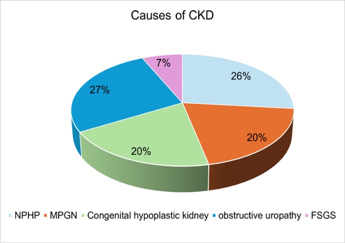
Figure 1. Etiology of Chronic Kidney Disease
The comparison of clinical characteristics measured before and after dialysis sessions is shown in Table II. The mean pre-dialysis weight was followed by a significant decrease after the first and subsequent treatment sessions. The blood pressure measurements showed a notable reduction in mean SBP and DBP.
Table I: Demographic Information and Base Line Characteristics |
|
Variables |
n (%) or mean ± SD |
Age (years) |
|
Plea5-10 |
9(30%) |
11-15 |
21(70%) |
Gender |
|
Male |
16(53.3%) |
Female |
14(46.7%) |
Clinical Markers |
|
Weight |
25.39±3.79 |
Pre dialysis SBP |
150.67±16.06 |
Pre dialysis DBP |
92.6±9.9 |
Pre dialysis IVC diameter |
11.68±1.49 |
Hemodialysis frequency / week (HDF) |
|
Twice |
24(80%) |
Thrice |
6(20) |
Pre dialysis pleural effusion (PE) |
|
No |
4(13.3%) |
Yes |
26 (86.7) |
Pre dialysis pericardial effusion (PCE) |
|
No |
4(13.3%) |
Yes |
26(86.7%) |
Pre dialysis B lines |
|
No |
11(36.7%) |
Yes |
19(63.3%) |
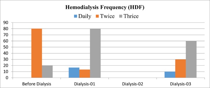
Figure 2 Prescription of Hemodialysis Frequency
Table 2 - Comparison of Clinical Characteristics Before and After Dialysis Sessions |
||||
Variable |
Before Dialysis Mean ± SD |
Dialysis-01 |
Dialysis-02 |
Dialysis-03 |
Weight (kg) |
25.39±3.79a |
23.96±3.73be |
25.11±3.67c |
23.83±3.61de |
Blood pressure (mmHg) |
||||
SBP |
150.67±16.06b |
135.37±11.47a |
143.73±16.65b |
143.73±16.65b |
DBP |
92.6±9.9b |
83.33±16.21a |
88.47±9.97ab |
86.67±9.94ab |
IVC diameter (mm) |
11.68±1.49b |
8.26±1.23a |
11.39±1.35b |
9.0±1.73ca |
HDF |
||||
Daily |
0(0%)a |
5(16.7%)a |
|
3(10%)b |
Twice |
24(80%)a |
1(3.3%)a |
|
9(30%)b |
Thrice |
6(20%)a |
24(80.%)a |
|
18(60%)b |
Pleural Effusion |
||||
No |
4(13.3%)a |
4(13.3%)a |
10(33.3%)b |
16(53.3%)c |
Yes |
26 (86.7%)a |
26(86.7%)a |
20(66.7%)b |
14(46.7%)c |
Pericardial Effusion |
||||
No |
4(13.3%)a |
11(36.7%)a |
22(73.3%)b |
29(96.7%)c |
Yes |
26(86.7%)a |
19(63.3%)a |
8(26.7%)b |
1(3.3%)c |
B lines |
||||
No |
11(36.7%)a |
11(36.7%)a |
20(66.7%)b |
27(90%)c |
Yes |
19(63.3%)a |
19(63.3%)a |
10(33.3%)b |
3(10.0%)c |
SBP: systolic blood pressure; DBP: diastolic blood pressure; IVC: inferior vena cava; HDF: Hemodialysis frequency |
||||
Previously, about 80% of patients underwent twice weekly dialysis and 20% were advised three sessions per week. The dialysis prescription was adjusted to daily sessions in 16.7% children, 3.3% received treatment twice a week, and 80% underwent dialysis thrice a week (Figure 2). These findings illustrate the dynamic changes in clinical characteristics throughout the dialysis sessions, highlighting the impact of the treatment on various health parameters.
Table 3 Patients with Persistent Fluid Overload Despite Third Dialysis Treatment (n = 6) |
|||
Variables |
a (n=3) |
b (n=3) |
p-value |
IVC |
9±1.73 |
7.9±0.46 |
.250 |
WT |
25.43±2.23 |
24.4±1.84 |
.250 |
SBP |
126.67±5.77 |
123.33±5.77 |
.250 |
DBP |
83.33±5.77 |
83.33±5.77 |
1.00 |
Pleural effusion (PE) |
|||
No |
2(66.7%) |
3(100%) |
|
Yes |
1(33.3%) |
|
|
Pericardial effusion (PCE) |
3(100%) |
|
|
No |
3(100%) |
|
|
Yes |
|
|
|
B lines |
|||
No |
3(100%) |
3(100%) |
|
Frequency HDF |
|||
Daily |
3(100%) |
3(100%) |
|
Table III displays the characteristics of the remaining 6 patients with persistent fluid overload despite repeated three dialysis sessions. They were compared as two groups 'a' and 'b'. The key findings and corresponding p-values are outlined regarding weight, IVC diameter, and blood pressure measurements along with radiological presence of pleural / pericardial effusion and B lines. Although the p-values were not suggestive of statistical significance, the HDF in both the groups was prescribed as daily hemodialysis.
B-lines exhibit a robust correlation with extravascular lung water which is a significant component of fluid volume.11 Several studies have identified a connection between B-line scores and weight fluctuations during the dialysis period.12,13The effectiveness of lung ultrasound in detecting changes in weight has been validated in pediatric studies.8 Numerous studies have explored B-line images.1,10,15,19 A study conducted in Egypt reports statistically significant decrease in the number of B-lines following dialysis treatment (p-value < 0.001) 20 which is consistent with findings in our study. To prevent biased measurements, we excluded patients with heart failure and active lung infection. Similarly, research by Torino et al 6 emphasized the importance of presence of B-lines seen on ultrasound in assessing fluid overload, highlighting its superiority over chest auscultation. Additionally, Allinovi and coworkers concluded that lung ultrasound is a more precise method for detecting subclinical fluid overload.8 We employed lung ultrasound and inferior vena cava (IVC) diameter measurements to adjust hemodialysis frequency as part of our treatment modification strategy. Pre- and post-dialysis sonographic measurements of IVC in our patients revealed a considerable decrease in diameter comparable to the findings with a study conducted in Egyptian pediatric population, which also reported a marked reduction in the IVC diameter after dialysis treatment (p-value < 0.001).20 Moreover, Torterüe et al14 observed a notable difference in IVC diameter measurements between pre- and post-dialysis sessions while evaluating fluid status in individuals on maintenance hemodialysis. As IVC diameter provides insight into intravascular filling grade and may be less influenced by volume changes during dialysis, it does have limitations, including inadequate visualization.9 Achieving and sustaining the appropriate dry weight proves to be quite challenging due to the absence of a clinical gold standard, but it is essential for ensuring dialysis adequacy in children, thereby preventing hypo- and hypertension during or after dialysis.15 In our study, a notable decrease in body weight of children was observed - 25.39 to 23.23 kg pre- and post-dialysis, respectively. These findings are similar to the results reported by Saadoon et al 2 which showed a significant decrease in body weight from 32.27 kg before dialysis to 30.58 kg after dialysis along with a significant decrease in systolic and diastolic BP measurements. Another new modality to assess the volume status in dialysis patients is bioimpedance approach. Ahmad et al found the use of bioimpedance spectroscopy (BIS) to be more sensitive than using the clinical parameters and IVC diameter.20 Contrary to this, another study concluded that IVC diameter measurement can represent intravascular volume measurement and it has significant advantage over BIS.18
Lung ultrasound presents a noninvasive, radiation-free and cost-effective method for evaluating fluid volume changes in pediatric dialysis patients, particularly through the measurement of inferior vena cava diameter and B-lines. This modern modality performed at the patients’ bedside enables adjustments to dialysis prescriptions based on the acquired information. Moreover, sequential lung scans can aid in revising dialysis frequencies to attain the desired dry weight.
Limitations: The sample size was small and the radiologist was not blinded to the pre- and post-dialysis ultrasound scans which could have introduced a repeated-measures bias.
- Razaq CM, Ijaz I, Bint-e-Mansoor K, Mehmood K, Tariq A, Abbas F. Volume Assessment in Pediatric Hemodialysis Using Lung Ultrasonography. Annals of King Edward Medical University. 2022 Aug 4;28(2):193-9. https://doi.org/10.21649/akemu.v28i2.5091
- Saadoon HASM, Youssef DYM, Ali A-SA, Neemat-Allah MAAJTEJoHM. Dry Weight Assessment in Children on Regular Hemodialysis with Special Relation Between Acute and Chronic Renal Failure. 2021;85(2):3953-9.
- Fu Q, Chen Z, Fan J, Ling C, Wang X, Liu X, et al. Lung ultrasound methods for assessing fluid volume change and monitoring dry weight in pediatric hemodialysis patients. 2021;36(4):969-76. https://doi.org/10.1007/s00467-020-04735-9
- Covic A, Siriopol D, Voroneanu LJAJoKD. Use of lung ultrasound for the assessment of volume status in CKD. 2018;71(3):412-22. https://doi.org/10.1053/j.ajkd.2017.10.009
- Kim HR, Bae HJ, Jeon JW, Ham YR, Na KR, Lee KW, et al.. A novel approach to dry weight adjustments for dialysis patients using machine learning. Plos one. 2021 Apr 23;16(4):e0250467. https://doi.org/10.1371/journal.pone.0250467
- Torino C, Gargani L, Sicari R, Letachowicz K, Ekart R, Fliser D, et al. The agreement between auscultation and lung ultrasound in hemodialysis patients: the LUST study. 2016;11(11):2005-11. https://doi.org/10.2215/cjn.03890416
- Al-Saray MZ, Ali A. Lung Ultrasound and Caval Indices to Assess Volume Status in Maintenance Hemodialysis Patients. POCUS journal. 2023;8(1):52. https://doi.org/10.24908/pocus.v8i1.15802
- Allinovi M, Saleem M, Romagnani P, Nazerian P, Hayes WJNDT. Lung ultrasound: a novel technique for detecting fluid overload in children on dialysis. 2017;32(3):541-7. https://doi.org/10.1093/ndt/gfw037
- Kaptein EM, Kaptein MJ. Inferior vena cava ultrasound and other techniques for assessment of intravascular and extravascular volume: an update. Clinical Kidney Journal. 2023 Nov;16(11):1861-77. https://doi.org/10.1093/ckj/sfad156
- Hayes W, Paglialonga FJPN. Assessment and management of fluid overload in children on dialysis. 2019;34(2):233-42. https://doi.org/10.1007/s00467-018-3916-4
- Beaubien-Souligny W, Rhéaume M, Blondin M-C, El-Barnachawy S, Fortier A, Éthier J, et al. A simplified approach to extravascular lung water assessment using point-of-care ultrasound in patients with end-stage chronic renal failure undergoing hemodialysis. 2018;45(1-3):79-87. https://doi.org/10.1159/000481768
- Fu Q, Chen Z, Fan J, Ling C, Wang X, Liu X, et al. Lung ultrasound methods for assessing fluid volume change and monitoring dry weight in pediatric hemodialysis patients. 2021;36:969-76. https://doi.org/10.1007/s00467-020-04735-9
- Loutradis C, Sarafidis PA, Ferro CJ, Zoccali CJNDT. Volume overload in hemodialysis: diagnosis, cardiovascular consequences, and management. 2021;36(12):2182-93. https://doi.org/10.1093/ndt/gfaa182
- Torterüe X, Dehoux L, Macher M-A, Niel O, Kwon T, Deschênes G, et al. Fluid status evaluation by inferior vena cava diameter and bioimpedance spectroscopy in pediatric chronic hemodialysis. 2017;18(1):1-7. https://doi.org/10.1186/s12882-017-0793-1
- Afifi W, Sweed E, Mohamed A, El-Gendy SJBJoAS. Lung Ultrasound: A Novel Technique for Detecting Fluid Overload in Children on Hemodialysis. 2022;7(5):95-100. https://dx.doi.org/10.21608/bjas.2022.244599
- Loutradis C, Sarafidis PA, Ekart R, Papadopoulos C, Sachpekidis V, Alexandrou ME, et al. The effect of dry-weight reduction guided by lung ultrasound on ambulatory blood pressure in hemodialysis patients: a randomized controlled trial. 2019;95(6):1505-13. https://doi.org/10.1016/j.kint.2019.02.018
- Arun Thomas E, Mohandas M, George JJHI. Comparison between clinical judgment and integrated lung and inferior vena cava ultrasonography for dry weight estimation in hemodialysis patients. 2019;23(4):494-503. https://doi.org/10.1111/hdi.12762
- Vitturi N, Dugo M, Soattin M, Simoni F, Maresca L, Zagatti R, et al. Lung ultrasound during hemodialysis: the role in the assessment of volume status. International urology and nephrology. 2014;46(1):169-74. https://doi.org/10.1007/s11255-013-0500-5
- Liang X-K, Li L-J, Wang X-H, Wang X-X, Wang Y-D, Xu Z-FJUiM, et al. Role of lung ultrasound in adjusting ultrafiltration volume in hemodialysis patients. 2019;45(3):732-40. https://doi.org/10.1016/j.ultrasmedbio.2018.10.025
- Ahmed EA, El-Mazary AA, Reyad AN, Abdel Wahhab MA, Moaness HM. Integrated lung and inferior vena cava ultrasonography for dry weight assessment in pediatric patients on regular hemodialysis: A single center study. Minia Journal of Medical Research. 2023 Jan 1;34(1):265-70. https://doi.org/10.21608/mjmr.2023.183994.1268

An Official Publication of
Islamabad Medical & Dental College
Vol 13 No (Suppl.)
Address of Correspondence
Noor ul ain Anwar
Email:
noorfjite@gmail.com
Authors’ Contribution: 1,2Conception; Literature research; manuscript design and drafting; 3,4 Critical analysis and manuscript review; 5,6Data analysis; Manuscript Editing.
Cite this article. Anwar A, Akhtar N, Perveen S, Sheikh R, Anjum MA, Saud S. Utilizing Integrated Lung and Inferior Vena Cava Ultrasound for Dry Weight Assessment - Insights from a Single-Center Experience. J Islamabad Med Dental Coll. 2024; 13i(Suppl.): 506-511 DOI: https://doi.org/10.35787/jimdc.
v13i(Suppl.).1202