Detection of Glioblastoma on Spectroscopy with Histopathology as Gold Standard
Ahson Ahmad1, Muhammad Mobeen2, Beenish Suhail3, Farheen Raza1, Muhammad Nauman Malik2, Sara Shahid1
1Department of Radiology, Shifa International Hospital Islamabad; 2Department of Radiology, Pakistan Institute of Medical Sciences, Islamabad; 3Department of Radiology, Akbar Niazi Teaching Hospital, Islamabad
Objective: The aim of this study was to ascertain the diagnostic accuracy of magnetic resonance spectroscopy (MRS) in the diagnosis of glioblastoma, using histopathology as the gold standard.
Methodology: In total 83 participants were engaged in this six-month cross-sectional descriptive study, carried out at the Department of Radiology, Shifa International Hospital in Islamabad. For each of these individuals, a single voxel approach was used for MR spectroscopy. To locate the lesion, post-contrast conventional MR imaging was first performed. A voxel was then placed on the region of interest. The pathology department at Shifa International Hospital in Islamabad received specimens from patients undergoing intracranial biopsies for histopathological investigation. The results of the MRS were then compared with the histopathology report.
Results: The average age of patients was 52.2±12.6 years with 34 females (51%), and 49 males (59%). The average length of the illness was 2.0±1.7 years, and the average lesion size was 54.3±26.9 mm. The results of MR spectroscopy diagnostic accuracy in the diagnosis of glioblastoma were 91.5%, with sensitivity, specificity, positive predictive value and negative predictive value of 90.7%, 94.4%, 98.3%, and 73.9% respectively.
Conclusion: Magnetic resonance spectroscopy is a valuable tool for glioblastoma diagnosis.
Keywords: Diagnostic Accuracy, Glioblastoma, Histopathology, MR spectroscopy, Sensitivity, Specificity.
About eighty-one percent of malignant brain lesions are gliomas, the most prevalent primary intracranial lesion.1 Of all gliomas, glioblastoma is the most frequent (45%) and has a 5% 5-year survival rate.1 Grade IV Astrocytoma, another name for glioblastoma, was previously referred to as glioblastoma multiforme. Tissue diagnosis and histological grading are currently the gold standards for the clinical care of brain tumors and they have a direct impact on patient survival rate.2,3 However, there are certain disadvantages to histopathology. The first possibility is sampling error, an inevitable mistake that could lead to a false diagnosis. Second, histopathology cannot reach any tumor remnants.4
Magnetic resonance spectroscopy is a non-invasive method of measuring tissue metabolite levels. MR spectroscopy makes it possible to compare the chemical composition of normal brain tissue and aberrant tumor tissue. Glioblastomas and other intracranial lesions can now be accurately diagnosed due to the advancement of magnetic resonance imaging in recent years. However, a lot of brain cancers lead to differential diagnosis because of MRI's great sensitivity and limited specificity. Magnetic resonance spectroscopy has been added to magnetic resonance imaging as an additional approach to increase its specificity.5 While MRI is used to visualize anatomical structures, MRS adds the ‘spectroscopy’ element to MRI resulting in determination of the underlying chemical composition within tissues and is thus crucial in distinguishing similar tumor types based on their biochemical composition.6 In terms of identifying tumors, MRS has a 77% sensitivity and a 84.2% specificity in distinguishing glioma grades.7
For the ultimate diagnosis of glioblastoma, doctors still prefer biopsies, however, the MRS can help limit the differential and avoid unnecessary biopsies. Thus, evaluation of MRS's effectiveness and prospective application as a less intrusive, less expensive, and preferred substitute for biopsy are necessary. The objective of this study is to evaluate MRS's diagnostic efficacy and accuracy in identifying glioblastoma tumors relative to histopathology, the gold standard.
This cross-sectional study was conducted in the Department of Radiology, Shifa International Hospital in Islamabad, from December 2020 to June 2021). Before conducting this study, an ethical approval was obtained from the Institutional Review Board and Ethics Committee, Shifa International Hospital. The research involved the enrollment of 83 patients in total. The sample size was determined by utilizing the WHO calculator to compute the sensitivity and specificity, taking into account the 45% prevalence of gliomas, the 91.7% sensitivity of MRS for glioblastoma, the 94.3% specificity, and the 9% prevision level (8). A non-probability, consecutive sampling strategy was used in the investigation. All patients with suspicion of Glioblastoma on MR (as per-operational definition) from 30-70 years, both genders were included in the study. Patients who lost follow-up, patients with history of previous brain surgery, with MRS incompatible prosthesis or cardiac pacemaker holders, patients with claustrophobia history, pregnant and breastfeeding females and patients not willing to undergo biopsy of intracranial lesions were excluded from the study.
Patients with suspicion of glioblastoma on MRS, who met the inclusion/exclusion criteria were referred from the neurosurgery department, neurology departments and outpatient department were selected for the study. For each of these individuals, a single voxel approach was used for MR spectroscopy. To locate the lesion, post-contrast conventional MR imaging was first performed. A voxel was then placed on the region of interest. Following water suppression, the results were acquired with settings comprising TE (echo time) and TR (repetition time) of 135 and 1500, respectively, using a point-resolved spectroscopy (PRESS) approach for localization. Consultant radiologists (having completed at least five years of post-fellowship training) evaluated all of the pictures to determine whether or not the signs of glioblastoma were present (according to the operational definition). The pathology department at Shifa International Hospital in Islamabad received specimens from patients undergoing intracranial biopsies for histopathological investigation.
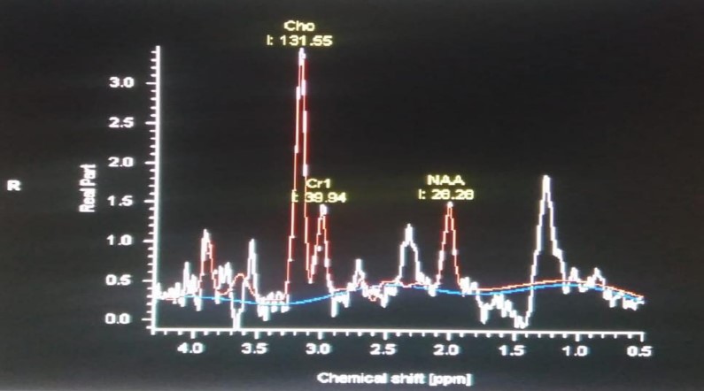
Figure 1: Detection of various chemicals on the spectroscopic spectrum
In total 83 patients were included in the study. Their age range was from 30 to 70 years with mean age was 52.2±12.6 years. 34 females (51%), and 49 males (59%) were present. The mean duration of disease was 2.0±1.7 years, and the mean lesion size was 54.3±26.9 mm. Sensitivity, specificity, positive predictive value, negative predictive value, and diagnostic accuracy of MR spectroscopy in the diagnosis of glioblastoma were 90.7%, 94.4%, 98.3%, and 91.5%, respectively.
Table I: Comparison of Magnetic resonance Spectroscopy |
|||
Magnetic resonance Spectroscopy |
Histopathology |
Total |
|
Positive |
Negative |
||
Positive |
59 (TP) |
1 (FP) |
60 |
Negative |
6 (FN) |
17 (TN) |
23 |
Total |
65 |
18 |
83 |
Table I shows diagnostic accuracy of MRS compared to Histopathology for glioblastoma. As shown in the table 59 patients were True positive, one False positive, six were False Negative and 17 cases were True Negative. The ROC curve analysis was carried out and the Area under the curve was observed as 0.91 as shown in Figure 2.
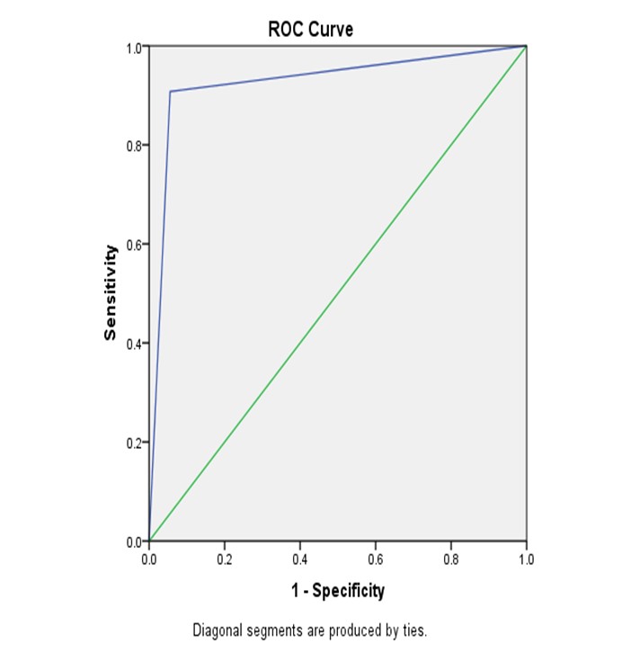
Figure 2: Area under the curve (AUC) is calculated out to be 0.91 as shown by the ROC curve analysis.
Glioblastomas multiforme (GBM) is one of the most common brain tumors (54% of gliomas) in adults. Grade IV glioma (according to the World Health Organization) is the most fatal brain tumor.8 Its one-year survival rate is 30%,9 while its five-year survival rate is 7.2%.10 Histological grade is a very important predictor of malignant gliomas.11 Stereotactic needle biopsy is a frequently used method to get a tissue diagnosis of brain lesions.12 Heterogeneity in gliomas may comprise of a complex mixture of malignant, necrotic, inflammatory and benign tissues.
Table II: stratification of diagnostic efficiencies with regard to age, gender, duration of disease and size of lesion |
||||||||
Characteristic |
Age (30-50 years) |
Age (51-70 years) |
Gender (Male) |
Gender (Female) |
Duration of Disease (≤ 4 year) |
Duration of Disease (> 4 year) |
Size of lesion |
Size of lesion (> 50mm) |
Sensitivity |
85.2% |
96.7% |
92.6% |
87.5% |
91.2% |
87.5% |
96.7% |
85.2% |
Specificity |
100% |
92.3% |
100% |
90% |
93.3% |
100% |
90% |
100% |
Positive Predictive Value |
100% |
96.7% |
100% |
95.4% |
98.1% |
100% |
96.7% |
100% |
Negative Predictive Value |
50% |
92.3% |
72.3% |
75% |
73.6% |
75% |
90% |
61.5% |
Accuracy |
87.1% |
95.4% |
93.8% |
88.2% |
91.6% |
90.9% |
95.1% |
88.1% |
Glioma biopsies taken in different places may therefore result in different histological grading. Stereotactic needle biopsy and glioma excision are further complicated due to concerns about possible neurologic effects from disruption in motor and expressive brain regions.13 Therefore, taking into account this issue in prognosis, it is important to develop non-invasive means for detection of a suitable place with possibly the highest histological grading before a biopsy or resection. While increasing Cho is a sign of rapid cell turnover and is elevated in disorders with enhanced cellular proliferation, such as malignant glioma, reduced NAA level indicates neuronal death.14,15 According to studies, neoplasms could be identified with a sensitivity of 77% and specificity of 84.2% at a cutoff value of 1.6 (NAA/Cr ratio of 1.7421), and high-grade gliomas could be distinguished from low-grade gliomas with a sensitivity of 74.2% and specificity of 62.5%.16,17 With a median lifespan of 6.9 months, glioblastoma (GBM), the most prevalent and deadly primary brain tumor in adults. It has significant molecular characteristics that makes it unavoidable to progress following standard therapy.18 Despite the severe standard first line treatment consisting of radiation and temozolomide usage (RT and TMZ), tumor recurrence occurs in nearly all patients.19However, GBM recurrence often shows radiologic characteristics similar to therapy related changes including a pseudoprogression (PsP) on conventional MRI. As a result, early and precise diagnosis of GBM relapse is essential. This is particularly important because of the growing number of potentially beneficial drugs being studied for salvage treatment. When compared to typical structural MRI techniques,
advanced imaging methods such as proton MR spectroscopy (MRS) and diffusion-weighted imaging (DWI) with apparent diffusion coefficient (ADC) mapping provide a deeper, non-invasive insight into the understanding of brain lesions. It increases the level of specificity in diagnostic imaging.20
Precise evaluation of the response to the initial treatment is crucial, and the most important prerequisite for an effective salvage therapy is the fast and correct detection of a tumor relapse. The current RANO (Response Assessment in Neuro-Oncology) criteria,21 already reflect the difficulties in distinguishing between GBM recurrence and treatment-related effects, such as pseudo progression from combined RT and TMZ therapy or pseudo response from angiogenesis inhibitors.22 Our results are further aligned with two more investigations conducted by Sahu et al and Amin et al.23,24 The MRS seems to be a promising method that can be used to increase the diagnostic accuracy of the brain tumor imaging procedure besides the widely used structural MRI. The findings of the current study revealed that MRS has very high sensitivity (90.7%) and specificity (94.5%) in diagnosing glioblastoma. The diagnostic accuracy was 91.5%, with a positive predictive value (PPV) of 98.3% and a negative predictive value (NPV) of 73.9%. These results align with those reported by Alshammari et al.25
The gold standard for diagnosis of Glioblastoma is Histopathology, however there are certain limitations to it. MRS offers a minimally invasive, spectroscopic alternative to biopsies. In this study we tried to establish the diagnostic efficiency of MRS compared to the gold standard i.e., histopathology. Our results indicate that MRS can offer a minimally invasive reliable alternative to histopathology and should be used in clinical settings.
Limitations: The first limitation is the limited generalizability of the results due to a small sample size from one tertiary care center in Pakistan. Moreover, the study was conducted in a short span of 6 months which can affect the acquisition of long-term trends in patient characteristics.
Disclaimer: The current manuscript is part of Dr. Ahson’s MS thesis project.
- Hanif F, Muzaffar K, Perveen K, Malhi SM, Simjee SU. Glioblastoma Multiforme: A Review of its Epidemiology and Pathogenesis through Clinical Presentation and Treatment. Asian Pac J Cancer Prev APJCP. 2017;18(1):3–9. https://doi.org/10.22034/apjcp.2017.18.1.3
- Gao H, Jiang X. Progress on the diagnosis and evaluation of brain tumors. Cancer Imaging. 2013 Dec 11;13(4):466–81. https://doi.org/10.1102/1470-7330.2013.0039
- Tandel GS, Balestrieri A, Jujaray T, Khanna NN, Saba L, Suri JS. Multiclass magnetic resonance imaging brain tumor classification using artificial intelligence paradigm. Comput Biol Med. 2020 Jul 1; 122:103804. https://doi.org/10.1016/j.compbiomed.2020.103804
- Wang W, Hu Y, Lu P, Li Y, Chen Y, Tian M, et al. Evaluation of the diagnostic performance of magnetic resonance spectroscopy in brain tumors: a systematic review and meta-analysis. PloS One. 2014;9(11): e112577. https://doi.org/10.1371/journal.pone.0112577
- Kaddah RO, Khalil ME. Malignant focal brain lesions. Value of MRS tumour biomarkers in preoperative prediction of grades of malignancy. Egypt J Radiol Nucl Med. 2014 Dec 1;45(4):1201–8. https://doi.org/10.1371/journal.pone.0112577
- Mckee AC, Daneshvar DH. Chapter 4 - The neuropathology of traumatic brain injury. In: Grafman J, Salazar AM, editors. Handbook of Clinical Neurology [Internet]. Elsevier; 2015 [cited 2024 Sep 26]. p. 45–66. (Traumatic Brain Injury, Part I; vol. 127). https://doi.org/10.1016/b978-0-444-52892-6.00004-0
- Rafique Z, Awan MW, Iqbal S, Usmani NN, Kamal MM, Arshad W, et al. Diagnostic Accuracy of Magnetic Resonance Spectroscopy in Predicting the Grade of Glioma Keeping Histopathology as the Gold Standard. Cureus. 14(2):e22056. https://doi.org/10.7759/cureus.22056
- Ostrom QT, Bauchet L, Davis FG, Deltour I, Fisher JL, Langer CE, et al. Editor’s choice: The epidemiology of glioma in adults: a “state of the science” review. Neuro-Oncol. 2014 Jul;16(7):896. https://doi.org/10.1093/neuonc/nou087
- Mohammed S, Dinesan M, Ajayakumar T. Survival and quality of life analysis in glioblastoma multiforme with adjuvant chemoradiotherapy: a retrospective study. Rep Pract Oncol Radiother. 2022 Dec 29;27(6):1026–36. https://doi.org/10.5603/rpor.a2022.0113
- Wu W, Klockow JL, Zhang M, Lafortune F, Chang E, Jin L, et al. Glioblastoma Multiforme (GBM): An overview of current therapies and mechanisms of resistance. Pharmacol Res. 2021 Sep; 171:105780. https://doi.org/10.1016/j.phrs.2021.105780
- Śledzińska P, Bebyn MG, Furtak J, Kowalewski J, Lewandowska MA. Prognostic and Predictive Biomarkers in Gliomas. Int J Mol Sci. 2021 Sep 26;22(19):10373. https://doi.org/10.3390/ijms221910373
- Trojanowski P, Jarosz B, Szczepanek D. The diagnostic quality of needle brain biopsy specimens obtained with different sampling methods – Experimental study. Sci Rep [Internet]. 2019 [cited 2024 Mar 4];9. https://doi.org/10.1038/s41598-019-44622-4
- Chen CC, White NS, Farid N, Kobalka P, Hansen L, Pearn M, et al. pre-operative cellularity mapping and intra-MRI surgery: potential for improving neurosurgical biopsies. Expert Rev Med Devices. 2015 Jan;12(1):1–5. https://doi.org/10.1586/17434440.2015.975118
- McKnight TR, von dem Bussche MH, Vigneron DB, Lu Y, Berger MS, McDermott MW, et al. Histopathological validation of a three-dimensional magnetic resonance spectroscopy index as a predictor of tumor presence. J Neurosurg. 2002 Oct;97(4):794–802. https://doi.org/10.3171/jns.2002.97.4.0794
- Dowling C, Bollen AW, Noworolski SM, McDermott MW, Barbaro NM, Day MR, et al. Preoperative proton MR spectroscopic imaging of brain tumors: correlation with histopathologic analysis of resection specimens. AJNR Am J Neuroradiol. 2001 Apr;22(4):604–12. https://pmc.ncbi.nlm.nih.gov/articles/PMC7976037/
- Law M, Yang S, Wang H, Babb JS, Johnson G, Cha S, et al. Glioma grading: sensitivity, specificity, and predictive values of perfusion MR imaging and proton MR spectroscopic imaging compared with conventional MR imaging. AJNR Am J Neuroradiol. 2003;24(10):1989–98. https://pmc.ncbi.nlm.nih.gov/articles/PMC8148904/
- Hourani R, Brant LJ, Rizk T, Weingart JD, Barker PB, Horská A. Can proton MR spectroscopic and perfusion imaging differentiate between neoplastic and nonneoplastic brain lesions in adults? AJNR Am J Neuroradiol. 2008 Feb;29(2):366–72. https://doi.org/10.3174/ajnr.a0810
- Dusek L, Muzik J, Maluskova D, Májek O, Pavlík T, Koptíková J, et al. Cancer incidence and mortality in the Czech Republic. Klin Onkol Cas Ceske Slov Onkol Spolecnosti. 2014;27(6):406–23. https://doi.org/10.14735/amko2014406
- Stupp R, Mason WP, van den Bent MJ, Weller M, Fisher B, Taphoorn MJB, et al. Radiotherapy plus concomitant and adjuvant temozolomide for glioblastoma. N Engl J Med. 2005 Mar 10;352(10):987–96. https://doi.org/10.1056/nejmoa043330
- Ahmed R, Oborski MJ, Hwang M, Lieberman FS, Mountz JM. Malignant gliomas: current perspectives in diagnosis, treatment, and early response assessment using advanced quantitative imaging methods. Cancer Manag Res. 2014;6:149. https://doi.org/10.2147/cmar.s54726
- Wen PY, Macdonald DR, Reardon DA, Cloughesy TF, Sorensen AG, Galanis E, et al. Updated response assessment criteria for high-grade gliomas: response assessment in neuro-oncology working group. J Clin Oncol Off J Am Soc Clin Oncol. 2010 Apr 10;28(11):1963–72. https://doi.org/10.1200/jco.2009.26.3541
- Lc H da C, I R, Rc D, El G, Ag S. Pseudoprogression and pseudoresponse: imaging challenges in the assessment of posttreatment glioma. AJNR Am J Neuroradiol [Internet]. 2011 Dec [cited 2024 Mar 4];32(11). https://doi.org/10.3174/ajnr.a2397
- Amin U, Ahmad A, Ather S, Ahmed A. Diagnostic Accuracy of Magnetic Resonance Spectroscopy in Predicting the Grade of Glioma Keeping Histopathology as the Gold Standard. PJR [Internet]. 2019 Mar 30 [cited 2024 Mar 4];29(1).
- Sahu A, Mathew R, Ashtekar R, Dasgupta A, Puranik A, Mahajan A, et al. The complementary role of MRI and FET PET in high grade gliomas to differentiate recurrence from radionecrosis. Front Nucl Med [Internet]. 2023 [cited 2024 Mar 4];3. https://doi.org/10.3389/fnume.2023.1040998
- Alshammari QT, Salih M, Gameraddin M, Yousef M, Abdelmalik B, Loaz O. Accuracy of Magnetic Resonance Spectroscopy in Discrimination of Neoplastic and Non-Neoplastic Brain Lesions. Curr Med Imaging. 2021 Jan 28;17(7):904–10. https://doi.org/10.2174/1573405617666210224112808

An Official Publication of
Islamabad Medical & Dental College
Vol 13 No (Suppl.)
Address of Correspondence
Muhammad Mobeen
Email:
mobeen916@yahoo.com
Authors’ Contribution: 1,2Conception; Literature research; manuscript design and drafting; 3,4 Critical analysis and manuscript review; 5,6Data analysis; Manuscript Editing.
Cite this article. Ahmad A, Mobeen M, Suhail H, Raza F Malik MN, Shahid S. Detection of Glioblastoma on Spectroscopy with Histopathology as Gold StandardJ Islamabad Med Dental Coll. 2024; 13i(Suppl.): 512-517
DOI: https://doi.org/10.35787/jimdc.
v13i(Suppl.).1238