An Extensive Infiltrative Diffuse Tenosynovial Giant Cell Tumor in Both Compartments of the Forearm: A Case Report
Safdar Ali Khan1, Muhammad Suhail Arif1, Abdullah Safdar1, Hamza Safdar2
1Department of Orthopedic Surgery, Fatima Memorial Hospital (FMH)/College of Medicine and Dentistry, Lahore, 2Department of Orthopedic Surgery, Ghurki Trust Teaching Hospital, Lahore
Diffuse tenosynovial giant cell tumors are relatively rare but second most common tumor effecting the synovial containing tissues. The study aim was to present a case involving a 40-year-old male patient with a complaint of a progressively enlarging swelling on the radial side of right wrist. On local examination, there was a bi-lobed swelling 7×9 and 6×5 cm on the dorsolateral and anterolateral aspect of right wrist respectively. Through Hennery’s modified approach tumor was exposed. It extended from the dorsolateral to the anteromedial wrist encasing the flexors, extensor and abductor tendons of the thumb and radial neurovascular bundle and adjacent wrist and finger flexors. The tumor surrounding the tendons was carefully removed, and carpal tunnel release was performed to address the tingling sensation. The patient managed was having a lobulated diffuse type of tumor involving the both flexor and extensor compartments. Better outcomes, less morbidity and least chance of recurrence can be expected if the tumor had been operated earlier.
Keywords: Giant cell tumor of tendon sheath; Tendon entrapment; Tenosynovitis.
Tenosynovial giant cell tumors (TS-GCT) was considered as a malignant tumor. TS-GCT was found to occur more often in middle-aged women than in men.1 TS-GCTs are the second most common soft tissue tumors in the hand. It is a proliferative disorder originating from bursae, tendons, and synovium of joints.2 TS-GCT are divided into two types depending upon the area of origin; one is non tenosynovial and the other tenosynovial. Non tenosynovial GCTs are less common and exhibit a range of biological behaviors, from borderline to fully malignant disease. Whereas, Tenosynovial GCTs, also referred to as GCTs of the tendon sheath or pigmented villonodular synovitis (PVNS) are almost benign, with high tendency for local recurrence.3 Byers has classified TS-GCT according to the growth pattern into localized nodular type, which are mostly found in the hand and diffuse type, which are common around the joints and is notorious for its invasiveness, recurrence and malignant transformation.4 In this case report, our experience with a diffuse TS-GCT of the right forearm case which has involved the radial artery other than both flexor and extensor compartment of the forearm. Presence of this kind of lesions in such an unusual location can be misleading, diagnostically, and surgically difficult to excise. This study also briefly discusses the common clinical, histological, and radiological features observed in literature review.
After receiving approval from the institute's ethics committee and securing informed written consent, a 40-year-old male patient presented in outpatient department of Orthopedic at Fatima Memorial Hospital, Lahore in December 2023 with the complaint of progressively enlarging swelling on the radial side of his right wrist from the last 2.5 years. The patient first noticed a small, peanut-sized swelling on the dorsum of his wrist about one and a half years ago. As the lesion was initially very small, painless, and did not affect daily life, patient ignored the swelling until it became large enough, tender and cosmetically ugly. There was no history of trauma, fever, anorexia, weight loss or evening rise of temperature. There was on similar swelling in any other part of his body and all joints were normal. The patient had no systemic disease and no relevant family medical history. General and systemic physical examination was unremarkable. On local examination, there was a bi-lobed swelling 7×9 and 6×5 cm on the dorsolateral and anterolateral aspect of his right wrist respectively (Figure 1). It was non-pulsatile, normo-thermic, mild tender, firm and non- compressible. Swelling was irregular, non-trans-illuminant, non-mobile and non- adherent to overlying normal skin but attached to the underlying flexors and extensor tendons. Radial artery was palpable proximal to the swelling. His distal neurology was intact except patient had slight tingling in the median nerve dermatome. Wrist was deviated ulnar ward, and the mass slightly reduced the degree of radial deviation but was painless. The results of the routine laboratory blood tests, inflammatory markers and X-rays of right forearm and wrist were normal. Ultrasound report revealed a bilobed soft tissue lesion with minimal vascularity on the anterolateral and posterolateral surface of the wrist.
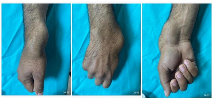
Figure 1: A mass located on the dorsolateral and anterolateral right wrist
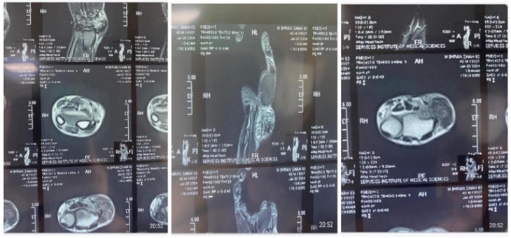
Figure 2: MRI images. The flexors and extensor tendons of thumb and index finger and abutting those of middle finger. Radial neurovascular completely encased by this mass.
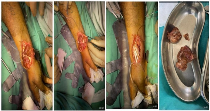
Figure 3: Preoperative & post operative identification of radial artery and excision of tumor.
Tenosynovial giant cell tumors (TS-GCT) are the second most common soft tissue tumors in the hand after the ganglion cyst.3 The female-to-male ratio is 3:2. However, unlike localized TS-GCT, diffuse type TS-GCT is notorious for bone erosion and adjacent soft tissue invasion.4 Diffuse type TS-GCT is locally aggressive and recurs in 40–60% of cases. It has a lot of comorbidity and often requires multiple surgical interventions. Histopathologic confirmation and definite classification of these tumours have important clinical implications.5 GCTS is commonly found in individuals aged 30 to 50 years. Clinical presentation of TS-GCT is variable and depends upon the site of origin. The first sign usually is a painless small nodule which enlarges slowly and progressively. It may eventually become painful, edematous and tender as it impinges on the surrounding anatomic structures.3 Clinical diagnosis is often challenging because of its unusual locations and nonspecific symptoms. Radiographs mostly show soft tissue shadow and are usually not very helpful but may show cortical erosions of the bone. Computed tomography and ultrasonography provide limited additional information. The definite diagnosis is only possible by immunochemistry, histologically or by MRI. Most TS-GCTs appear iso-intense compared to muscles on T1-WI imaging, with varying intensity on T2-WI imaging due to the presence of fluid, hemosiderin, lipid, hemorrhagic components, and fibrous tissue.6
An extensive TS-GCT of the forearm in this case report also exhibited aggressive invasion the flexors, extensor and abductor tendons of thumb adjacent wrist and finger flexors and encased radial neurovascular bundle. Marginal excision of the diffuse TS-GCT is the surgical treatment of choice but surgical management of local aggressive diffuse-type TS-GCT is challenged due to pathological tissue spread to the adjacent soft tissue and might be technically difficult to remove. Sometime excision of the involved skin and reconstruction of the entrapped tendons may be required. However, meticulous dissection and exploration are essential in patients with extensive disease to avoid seeding the healthy surrounding structures. Even with careful dissection in diffuse TS-GCT reported recurrence rate is very high in the range of 9-44%.7
As high risk of recurrence, other treatments such as radiotherapy may be considered. To avoid recurrence after surgery many studies have been done to see the effectiveness of radiotherapy. In one meta-analysis patients with TS-GCT, using perioperative radiotherapy shown lower rate of recurrence but evidence was of low quality.7 Despite these results, radiotherapy is not popular in young patients due to its long-term toxicity and subsequent morbidity like radiation fibrosis, tissue necrosis, skin changes, and radiation to secondary malignant transformation.6 This reason could not convince us to use this modality. To prevent recurrence certain chemotherapeutic agents that target colony-stimulating factor (CSF1), receptor like imatinib, nilotinib, emactuzumab and pexidartinib have been investigated (PLX3397).8
Ensuring the best outcome for the patient is crucial in cases like this. The lobulated diffuse type of tumor involving both the flexor and extensor compartment, which also incorporates the radial neurovascular bundle, requires thorough monitoring and a longer follow-up period due to its complexity. Acting promptly and operating on the tumor earlier can significantly improve the prognosis, leading to a better outcome, less morbidity, and the lowest chance of recurrence. Immediate attention and intervention can make a substantial fast recovery.
- Choi WS, Lee SK, Kim JY, Kim Y. Diffuse-Type Tenosynovial Giant Cell Tumor: What Are the Important Findings on the Initial and Follow-up MRI? Cancers. 2024;16(2):402. https://doi.org/10.3390/cancers16020402
- Kropivsek L, Pizem J, Mavcic B. Giant Cell Tumor of Bone Versus Tenosynovial Giant Cell Tumor-Similarities and Differences. Int J Surg Pathol 2022;30(6):596-605. https://doi.org/10.1177/10668969221076545
- Robert M, Farese H, Miossec P. Update on tenosynovial giant cell tumour, an inflammatory arthritis with neoplastic features. Front Immunol. 2022;13(2022):820046. https://doi.org/10.3389/fimmu.2022.820046
- Alves L, Leite MJ, Lima S, Torres T, Gutierres M. Localized tenosynovial giant cell tumor in the posterior compartment of the knee-A rare case report. J Orthop Rep. 2023;2(3):100184. https://doi.org/10.1016/j.jorep.2023.100184
- Spierenburg G, Suevos Ballesteros C, Stoel BC, Navas Cañete A, Gelderblom H, van de Sande MA, et al. MRI of diffuse-type tenosynovial giant cell tumour in the knee: a guide for diagnosis and treatment response assessment. Insights Imaging. 2023;14(1):22. https://doi.org/10.1186/s13244-023-01367-z
- Chelli Bouaziz M, Ladeb MF, Riahi H. Synovial Hemangioma. In Imaging of Synovial Tumors and Tumor-like Conditions. 2023:163-172. Cham: Springer International Publishing. https://doi.org/10.1007/174_2023_417
- Siegel M, Bode L, Sudkamp N, Kuhle J, Zwingmann J, Schmal H, et al. Treatment, recurrence rates and follow-up of Tenosynovial Giant Cell Tumor (TGCT) of the foot and ankle-A systematic review and meta-analysis. PLoS One. 2021;16(12): e0260795. https://doi.org/10.1371/journal.pone.0260795
- Benner B, Good L, Quiroga D, Schultz TE, Kassem M, Carson WE, et al. Pexidartinib, a novel small molecule CSF-1R inhibitor in use for tenosynovial giant cell tumour: a systematic review of pre-clinical and clinical development. Drug Des Devel Ther. 2020;14(2020):1693-704. https://doi.org/10.2147/DDDT.S253232

An Official Publication of
Islamabad Medical & Dental College
Volume 13 Issue 3
Case Report
Address of Correspondence
Safdar Ali Khan
Email:
drsafdaralikhan@gmail.com
Authors’ Contribution: 1,2Conception; Literature research; manuscript design and drafting; 3,4 Critical analysis and manuscript review; 5Data analysis; Manuscript Editing.
Cite this article. Khan SA, Arif MS, Safdar A, Safdar H. An Extensive Infiltrative Diffuse Tenosynovial Giant Cell Tumor in Both Compartments of the Forearm: A Case Report. J Islamabad Med Dental Coll. 2024; 13(3): 486-489.
DOI: https://doi.org/10.35787/jimdc.v13i3.1258