Reticulocyte Haemoglobin Equivalent a Useful Indicator of Iron Deficiency in Patients with Iron Deficiency Anaemia and Anaemia of Chronic Disease
Kehkashan Hassan1, Saleem Ahmed Khan2, Fahim Akhtar3, Sunita Tashfeen4, Samra Shahid5, Maham Arshad Malik5
1Department of Haematology, Armed Forces Institute of Pathology, Rawalpindi, Pakistan; 2National University of Medical Sciences (NUMS), Rawalpindi; 3Department of Haematology, AFIT, CMH Rawalpindi; 4Department of Pathology, Army Medical College, National University of Medical Sciences (NUMS), Rawalpindi, Pakistan / Pak Emirates Military Hospital, Rawalpindi, Pakistan; 5Department of Pathology, Army Medical College, National University of Medical Sciences (NUMS), Rawalpindi, Pakistan;
Objectives: To find an association between RET-He and iron deficiency (ID) and evaluate its usefulness in detection of iron deficiency in patients with iron deficiency anaemia (IDA) and anaemia of chronic disease (ACD).
Methodology: A total of 95 participants were selected and divided into three groups control, IDA and ACD after taking informed consent and obtaining ethical approval. Patients were selected according to WHO diagnostic criteria for IDA and ACD and subjected to testing for RET-He by using sysmex XN 3000 which quantifies haemoglobin content of reticulocytes via fluorescent flow cytometry. Blood counts were collected and analyzed by using SPSS version 22.
Results: Mean age of total population was 44.62 years and ranged from 16- 85 years. Anemia was significant in female population in the IDA group. Mean value of RET-He in CG was 31.35 ± 1.5 4.16pg, in IDA group was 20.6 ± 4.16 and in ACD group was 26.3 ± 4.9 pg. RET-He was significantly low in the IDA group and in ACD it was moderately low. RET-He detected ID in both groups and a significant p-value of ˂0.001 was obtained.
Conclusion: RET-He is a useful indicator of ID which is not affected by inflammation and readily detects iron deficiency.
Keywords: anaemia of chronic disease; flow cytometry; iron deficiency anaemia; reticulocyte haemoglobin equivalent.
IDA is the most common nutritional deficiency in the world that nearly affects 700 to 800 million people worldwide.1 A vast majority of Pakistani population suffers from IDA. Various laboratory investigations that are used routinely to diagnose IDA include evaluation of haemoglobin concentrations (Hb), mean corpuscular volume (MCV), haematocrit (Ht), mean corpuscular haemoglobin concentration (MCHC) and red cell distribution width (RDW). Other biochemical tests include serum iron levels (Fe), serum ferritin (sFt) concentration, total iron binding capacity (TIBC), transferrin saturation (TSAT), soluble transferrin receptor (sTFR) as well as bone
marrow biopsy.2 It is difficult to diagnose IDA by using conventional biomarkers of iron metabolism and red cell parameters. The characteristic morphological features of hypochromic and microcytic red blood cells (RBC) classically appear only after an advanced iron deficient erythropoiesis 3 Red cell parameters ( MCV, MCHC and RDW ) provide information about the availability of iron in mature red blood cells and are not considered sensitive parameters as early indicators of ID because of slow turnover of erythrocytes (120 days).4 Alternative biochemical tests currently in use for the diagnosis of IDA have certain limitations and are influenced by many other conditions. Serum iron is low in both IDA as well as in anaemia of chronic disease and its level remains variable during day depending on the intake of iron. The value of TSAT is also affected by iron and TIBC as its calculation is based upon Fe and TIBC. Serum ferritin is considered a good predictor of iron storage and low levels indicate deficiency of iron. However, it acts as an acute phase reactant and can be elevated due to infections, inflammatory conditions and malignancy.5 The gold standard in evaluating iron deficiency is the bone marrow assessment of iron stores but is usually not preferred because of its invasive nature.6
Anaemia of chronic disease, now known as anaemia of inflammation (AI) is considered as the second most common anaemia in the world after IDA. It is the most frequent anaemia in chronically ill and hospitalized patients affecting ˃1 billion individuals worldwide.7 It occurs in response to biochemical changes in body which are mediated by chronic infections, malignancy, acute or chronic inflammatory diseases, or autoimmune disorders.8 IDA and anemia of inflammation (AI) often coexist and it is challenging to differentiate them from each other in the setting of inflammation.9 The evaluation of the biochemical markers of iron metabolism demonstrates a weakness in the diagnosis of functional iron deficiency occurring in ACD which is a growing concern in public health. Therefore, a newer, noninvasive and more standardized parameter is needed which could readily detect ID, eliminate the need for iron studies and bone marrow examination and can help in discriminating IDA from pure ACD especially in patients presenting with both IDA and ACD for better clinical outcome. Late occurrence of changes in red blood cell indices has recently generated an interest in using parameters of reticulocytes available on modern haaematology analyzers based on the technology of fluorescent flow cytometry for iron estimation. By using the forward scattered light and the fluorescence signal the reticulocytes can be separated from mature red blood cells. RET-He gives direct measurement of haemoglobin content of reticulocytes and reflects the quality of newly synthesized cells over past 3-4 days. Automated blood count devices include a reticulocyte count channel which can quantify haemoglobin content of reticulocytes and provide information regarding current iron status. Reticulocytes remain in peripheral blood for 1-2 days and measurement of their haemoglobin content can be a good index of iron deficiency. RET-He has been identified as a reliable indicator of iron deficient and iron restricted erythropoiesis and is a direct measure of amount of haemoglobin in young red blood cells. A range of 28-36 pg is considered normal for Ret He. It has been found to be more sensitive than serum ferritin in diagnosing iron deficiency and unlike ferritin is not an acute phase reactant.
This cross-sectional study was conducted from July 2020 to December 2020 after approval from Institutional Ethical Review Board (No ERC/ID/83), at Army medical College, National University of Medical Sciences (NUMS) in collaboration with Pak Emirates Military Hospital (PEMH), Rawalpindi. After obtaining informed consent 95 patients were included in the study using non probability consecutive sampling technique. Sample size was calculated by WHO sample size calculator taking confidence interval of 95%, margin of error 5%, power of test 80% and using reference prevalence as 45.5% of anticipated population of IDA. Patients were classified into three groups, Control group (n=30), IDA group (n=35) and ACD group (n=30). Patients of either gender, both newly diagnosed and untreated, above 13 years of age presenting in outpatient department with anaemia were included in the study. Patients having anaemia due to all other causes including congenital, patients on iron therapy and having received blood transfusions were excluded from the study. Control group consisted of disease-free healthy individuals with normal haematological findings, serum ferritin and CRP levels. WHO criteria were adopted and anaemia was defined as a haemoglobin level of ˂12 g/dL for females and of ˂13 g/dL for males. IDA was defined biochemically using serum ferritin levels of ˂ 20ng/mL and keeping it as a gold standard for detection of iron deficiency within anaemia patient 10. In IDA patients’ inflammatory disease was excluded by performing CRP. ACD was defined with normal or raised serum ferritin levels, haemoglobin levels ˃8 g/dl, MCV ˃ 80 fL, CRP levels ˃ 6mg/dl and presence of a chronic disease11. Patients from ACD group were suffering from a diverse group of diseases including chronic infections, inflammatory and autoimmune conditions, chronic kidney disease and malignancy.
Patients who did not give consent, pregnant females, patients on iron and erythropoietin therapy or patients with any other form of anaemia like megaloblastic, aplastic, haemoglobinopathies and hereditary forms were excluded from the study. Participant’s information was kept confidential and was not shared with anyone outside the organization. Every study subject was allotted a code number. Data was not accessible to anyone outside the research team and there was no conflict of interest. Sample was collected after explaining the established procedural aspects and aim of the study to all participants. Patients for the study were selected from the population referred by physicians to PEMH, department of hematology for investigation of anaemia, strictly following the inclusion and exclusion criteria. 5ml of blood venous blood sample was drawn under sterile conditions from each patient. About 3ml was transferred to EDTA tube and stored at 4˚C until complete blood picture was performed. Remaining blood was transferred in gel/plain tube for serum ferritin estimation by using machine Immulite 2000 and for performing CRP on Advia.
In this study we used sysmex analyzer of series XN-3000 having a reticulocyte channel analysis feature and utilizing impedance technology and fluorescent flow cytometry. After running quality control procedures, EDTA anticoagulated samples from all patients were run within 6 hours of collection and blood counts were generated. Frequencies and percentages were calculated for qualitative variables. Mean and standard deviation were calculated for quantitative variables. One way ANOVA was used to compare means of various groups. Data was entered and analyzed by using Statistical Package for Social Sciences (SPSS) version 22. The p-value of ≤ 0.05 was considered as significant.
The total study population consisted of 95 participants who were classified into three groups Control, IDA and ACD according to the WHO adopted criteria for each group. The group wise distribution of all participants is shown in table I.
Table - I: Group categories of all participants (n=95) |
||
Category |
No of |
|
Control group (CG) |
30 |
|
Iron deficiency anaemia (IDA) |
35 |
|
Anaemia of chronic disease (ACD) |
Chronic kidney |
10 |
Infections |
10 |
|
Rheumatoid arthritis |
5 |
|
Malignancy |
5 |
|
Mean age of total population was 44.6 years. Minimum age was 16 and maximum was 85 years. Out of total population there were 40(42.10%) males and 55(57.89%) females with a M/F ratio of 0.7:1. Majority of the patients in the IDA group were females whereas in the ACD group males showed a higher prevalence. Mean age of participants in the CG was 44.36 ±15.5 years, in IDA group was 33.94±11.91years and in ACD group was 57.33 ±18.1 years. Gender distribution of all participants is represented in Figure 1.
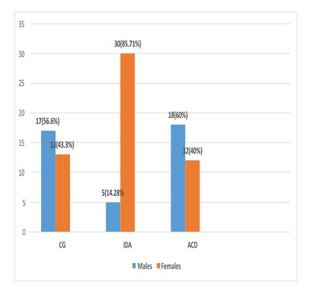
Figure.2: Gender distribution of all study participants (n=95)
Mean difference between three groups was identified and the results were compared between groups with regards to age, gender, RBC count, Hct and Hb levels, measurement of MCV, MCH, MCHC, RDW, Platelet count, biochemical findings of ferritin and CRP and estimation of Ret-He. Table - II represents the demographic features, haematological and biochemical findings of all groups.
Normal range considered for RET He was 28-36 pg. Our results remained significant with a p value of ˂0.001. Mean values 31.35 ± 1.5pg of RET-He in CG, 20.6 ± 4.1 6pg in IDA group and 26.3 ± 4.9 pg in ACD group were obtained which correlated well with our reference range. Red cell indices showed hypochromic microcytic erythrocytes in IDA group confirming IDA and normochromic normocytic erythrocytes in ACD group reflecting FID.
The mean RET-He values of all the groups are represented in table III whereas figure - 2 represents them graphically in the form of box and whisker graph.
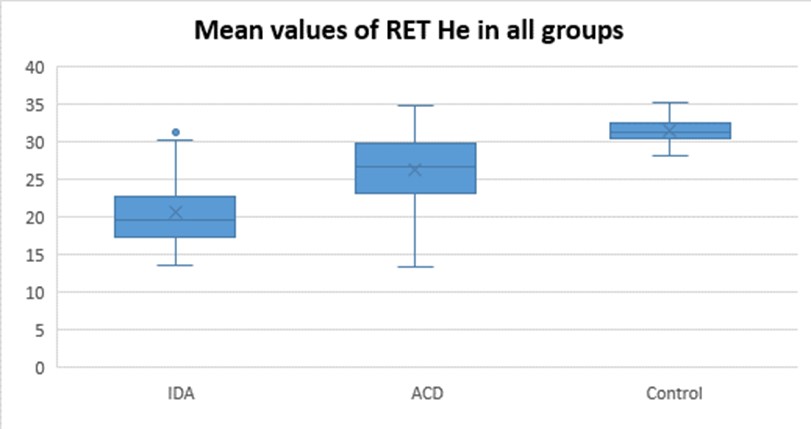
Figure - 2: Box and whisker graph showing mean values of RET He in all groups
Table - II: Haematological and biochemical findings of all groups (n=95) |
|||||
No |
Variable |
Control group (n=30) |
Iron deficiency |
Anaemia of chronic disease (n=30) |
P- value |
Mean ± SD |
Mean ± SD |
Mean ± SD |
|||
1 |
Red blood cell count |
4.90 ± 0.58 |
4.43 ± 0.74 |
3.65 ± 0.74 |
˂0.001 |
2 |
Hematocrit (Hct)(%) |
41.70 ± 4.0 |
30.51 ± 4.7 |
29.45 ± 4.40 |
˂0.001 |
3 |
Haemoglobin (Hb)(g/dL) |
14.32 ± 1.2 |
8.9 ± 1.4 |
9.5 ± 1.3 |
˂0.001 |
4 |
Mena corpuscular |
85.29 ± 3.7 |
69 ± 7.3 |
84 ± 6.6 |
˂0.001 |
5 |
Mean corupuscular |
29.0 ± 1.8 |
20.51 ± 3.0 |
27.49 ± 2.30 |
˂0.001 |
6 |
Mean corpuscular haemoglobin concentration |
33.72 ± 1.19 |
29.47 ± 1.8 |
32.96 ± 1.91 |
˂0.001 |
7 |
Platelet |
285.4 ± 71.5 |
366.11 ± 145 |
287 ± 91.6 |
˂0.004 |
8 |
Red cell distribution |
40.9 ± 3.6 |
45.57 ± 11.35 |
48.47 ± 11.07 |
˂0.001 |
9 |
C reactive protein |
1.036 ± 0.74 |
3.23 ± 1.01 |
120 ± 153.6 |
˂0.001 |
10 |
Ferritin(ng/mL) |
104 ± 40.63 |
8.4 ± 3.57 |
899.16 ± 1862 |
˂0.002 |
11 |
Reticulocyte |
31.35 ± 1.5 |
20.6 ± 4.16 |
26.3 ± 4.9 |
˂0.001 |
Table - III: Mean value of RET- He in all groups:
|
|||||||
RET – He |
Control group n=30 |
Iron deficiency anaemia |
Anaemia of chronic disease |
p- value |
|||
Mean ± SD |
Mean ± SD |
Mean ± SD |
˂0.001 |
||||
31.35 ± 1.5 |
20.60 ± 4.1 |
26.30 ± 4.9 |
|||||
Min |
28.20 |
Min |
13.60 |
Min |
13.40 |
˂0.001 |
|
Max |
35.10 |
Max |
31.20 |
Max |
34.70 |
˂0.001 |
|
Absolute and functional ID are the two major forms of Iron deficiency (ID) that can occur in an individual. Both these forms can appear either separately or in a combined form, and if not identified and left untreated, can result in iron deficient erythropoiesis and anemia.12 By using conventional laboratory tests it is not difficult to diagnose an uncomplicated ID as in an un-complexed setting, biochemical markers are considered reliable parameters to detect and diagnose ID. However, it becomes difficult to diagnose the ID or FID in patients with acute or chronic inflammation because the biochemical markers for metabolism of iron are greatly influenced and affected by acute phase reaction, and this is observed in anaemia associated with chronic conditions.13 Detection of ID is considered important in the management of patients with chronic disease because it cannot be corrected without parenteral iron therapy, and if ID is not present still it is important to identify because iron therapy has to be avoided as it will lead to iron overload which has serious implications.
Serum ferritin assay is the most widely used test and is considered essential in the assessment of all forms of iron restricted erythropoiesis including FID.14 However, ferritin largely loses its diagnostic value as an indicator of body iron stores in the setting of inflammation.15 TSAT is also unreliable and is considered less ideal for diagnosing ID in patients with ACD .16
The limitation of soluble transferrin receptor (sTfR) occurs in its lack of standardization.17 Moreover, it is relatively expensive and majority of the centres do not provide the facility. Use of ferritin index (FTI) is also limited as ferritin index is calculated from sTFR divided by logarithm of serum ferritin.18 In short, biochemical markers are considered less ideal for diagnosing ID particularly in ACD. Gold standard bone marrow has also been challenged because of its invasive nature and is avoided in diagnosis of ID. Therefore, an indicator superior than above all is still required for the evaluation of iron deficiency in different forms of anaemia. RET-He is the amount of haemoglobin in each reticulocyte reported in picograms, gives the real time information about the current supply of iron for erythropoiesis and is an early indicator of ID as compared to haemoglobin and hematocrit.
Our results were comparable to a number of studies. The role of RET-He has been validated by Mehta, et al., who reported RET-He, a better predictor of bone marrow iron store than ferritin in IDA. They determined its diagnostic accuracy by using bone marrow as a gold standard.19 Canals, et al., determined this parameter to be very useful in making rapid diagnosis. They determined its utility in thalassemia carriers and suggested that a direct diagnosis of IDA can be made in patients with mixed pattern anaemia.20
Rungngu et al., established a relationship between low RET-He and IDA and reported RET-He to be a useful screening tool in children for diagnosing IDA.4 Lourenco et al discovered its utility in detecting FID in ACD and differentiating ACD from ACD with coexistent ID.21 Sanyoto et al., reported strong association of RET- He with IDA and suggested its use as a screening tool for IDA.22 Tiwari, et al,. performed a study and demonstrated that it can be a useful marker for indicating ID in blood doners.23 Echardt et al., performed a study and found RET-He to be a useful marker for detection of ID in patients with chronic renal failure.24
RET-He is a useful marker for determining ID in general population. It is simple to measure and iron can be quantified quickly and cost effectively from a single EDTA blood tube during routine automated CBC plus reticulocyte count analysis and no additional blood sample and further chemical test is required. In patients with chronic disease where high ferritin levels secondary to inflammation may mask an underlying ID it can be used conveniently as it is not affected by inflammation.
Recommendations:
RET-He readily rules out iron deficient erythropoiesis however, future studies are required for validation of its efficacy and usefulness particularly in detection of coexistent IDA in ACD. Multi centred studies including different age groups and chronic inflammatory diseases should be carried out to assess the benefit of the information provided by RET-He. As we are still long way off from our goal to overcome IDA among vulnerable and hospitalized population, more attention is required to determine the iron status of patients with inflammatory conditions.
- Habib MA, Raynes-Greenow C, Soofi SB, Ali N, Nausheen S, Ahmed I, Bhutta ZA, Black KI. Prevalence and determinants of iron deficiency anemia among non-pregnant women of reproductive age in Pakistan. Asia Pacific journal of clinical nutrition. 2018 Jan;27(1):195-203. https://doi.org/10.6133/apjcn.042017.14
- Archer NM, Brugnara C. Diagnosis of iron-deficient states. Critical reviews in clinical laboratory sciences. 2015 Sep 3;52(5):256-72. https://doi.org/10.3109/10408363.2015.1038744
- Poffenroth C, Mabbett C, Kendrick C. The reticulocyte haemoglobin equivalent (RET_He) and laboratory screening for iron deficiency. New Zealand Journal of Medical Laboratory Science (N.Z.J.Med.Lab.Sci) 2017;71(3):120.
- Rungngu SLP, Wahani A, Mantik MF. Reticulocyte hemoglobin equivalent for diagnosing iron deficiency anemia in children. Paediatrica Indonesiana (Paediatr Indons). 2016;56(2):90-4. https://doi.org/10.14238/pi56.2.2016.90-4
- Uçar MA, Falay M, Dağdas S, Ceran F, Urlu SM, Özet G. The importance of RET-He in the diagnosis of iron deficiency and iron deficiency anemia and the evaluation of response to oral iron therapy. Journal of medical biochemistry (J.Med.Biochem). 2019;1. https://doi.org/10.2478/jomb-2018-0052
- Kılıç M, Özpınar A, Serteser M, Kilercik M, Serdar M. The effect of reticulocyte hemoglobin content on the diagnosis of iron deficiency anemia: A meta-analysis study. Journal of medical biochemistry. 2022 Feb 2;41(1):1. https://doi.org/10.5937/jomb0-31435
- Weiss G, Ganz T, Goodnough LT. Anemia of inflammation. Blood. 2019;133(1):40-50. https://doi.org/10.1182/blood-2018-06-856500
- Weimann A, Cremer M, Hernáiz-Driever P, Zimmermann M. Delta-He, Ret- He and a new diagnostic plot for differential diagnosis and therapy monitoring of patients suffering from various disease-specific types of anemia. Clin Lab. 2016;62(4):667-77. https://doi.org/10.7754/clin.lab.2015.150830
- Marques O, Weiss G, Muckenthaler MU. The role of iron in chronic inflammatory diseases: from mechanisms to treatment options in anemia of inflammation. Blood, The Journal of the American Society of Hematology. 2022;140(19):2011-23. https://doi.org/10.1182/blood.2021013472
- Enko D, Wagner H, Kriegshäuser G, Kimbacher C, Stolba R, Halwachs-Baumann G. Assessment of human iron status: a cross-sectional study comparing the clinical utility of different laboratory biomarkers and definitions of iron deficiency in daily practice. Clinical Biochemistry. 2015 Sep 1;48(13-14):891-6. https://doi.org/10.1016/j.clinbiochem.2015.05.008
- Nemeth E, Ganz T. Anemia of inflammation. Hematology/Oncology Clinics. 2014;28(4):671-81. https://doi.org/10.1016/j.hoc.2014.04.005
- Nairz M, Theurl I, Wolf D, Weiss G. Iron deficiency or anemia of inflammation? Wiener Medizinische Wochenschrift. 2016;166(13-14):411-23. https://doi.org/10.1007/s10354-016-0505-7
- Gelaw Y, Woldu B, Melku M. The Role of Reticulocyte Hemoglobin Content for Diagnosis of Iron Deficiency and Iron Deficiency Anemia, and Monitoring of Iron Therapy: a Literature Review. Clinical laboratory. 2019 Dec 1(12). https://doi.org/10.7754/clin.lab.2019.190315
- Fletcher A, Forbes A, Svenson N, Wayne Thomas D. Guideline for the laboratory diagnosis of iron deficiency in adults (excluding pregnancy) and children. British Journal of Haematology. 2022 Feb 1;196(3). https://doi.org/10.1111/bjh.17900 Dignass A, Farrag K, Stein J. Limitations of serum ferritin in diagnosing iron deficiency in inflammatory conditions. International journal of chronic diseases (Int.J.Chronic Dis). 2018;2018. https://doi.org/10.1155/2018/9394060
- Gupta DK, Choudhary RK, Sharma M, Saluja S, Gupta B. Role of soluble transferrin receptor and soluble transferrin receptor index in diagnosing iron deficiency anemia in patients with chronic kidney disease. Astrocyte. 2019;3(3):125.
- Pfeiffer CM, Looker AC. Laboratory methodologies for indicators of iron status: strengths, limitations, and analytical challenges. The American journal of clinical nutrition (AJCN). 2020;106(suppl_6):1606S-14S. https://doi.org/10.3945/ajcn.117.155887

An Official Publication of
Islamabad Medical & Dental College
Vol 13 No (Suppl.)
Address of Correspondence
Kehkashan Hassan
Email:
kehkashan_hassan@yahoo.com
Authors’ Contribution: 1,2Conception; Literature research; manuscript design and drafting; 3,4 Critical analysis and manuscript review; 5,6Data analysis; Manuscript Editing.
Cite this article. Hassan K, Khan SA, Akhtar F, Tashfeen S, Shahid S, Malik MA. Reticulocyte Haemoglobin Equivalent a Useful Indicator of Iron Deficiency in Patients with Iron Deficiency Anaemia and Anaemia of Chronic Disease J Islamabad Med Dental Coll. 2024;13i(Suppl.)
560-577. DOI: https://doi.org/10.35787/jimdc.
v13i(Suppl.).1314