Accuracy, Sensitivity and Specificity of Oral Brush Cytology in the Diagnosis of Oral Epithelial Lesions
Abdul Majid1, Bushra Sajid2, Kiran Shaikh2, Amin Fahim3, Farkhunda Nadeem4, Ghulam Shah Nizaman 4
1 Assistant Professor, Department of Pathology, Isra University Hyderabad
2 Lecturer, Department of Pathology, Isra University Hyderabad
3 Associate Professor, Department of Pathology, Isra University Hyderabad
4 Professor, Department of Pathology, Isra University Hyderabad
Background: Oral Squamous cell carcinoma (SCC) is highly prevalent in Pakistan than the rest of the world. Five-year survival rate is 80% if SCC is diagnosed at an early stage. The survival rate declines to 20% if diagnosed at a later stage. The objective of the present study was to find out the usefulness of oral brush cytology in detecting oral lesions.
Material and Methods:In the present prospective observational study, 88 samples were collected from Maxillofacial Surgery Out-Patient Department (OPD), Liaquat University Hospital and ISRA University Hospital from July 2015 to December 2015. After taking written informed consent, oral brush cytology was performed and stained with standard Papanicolaou (PAP) staining protocol. The biopsy of the patients was performed by standard protocols of oral biopsy and specimen preserved in 10% buffered formalin. Cross tabulation between diagnosis of brush cytology and biopsy of same patients was done and accuracy, sensitivity and specificity were calculated.
Results: According to the distribution of patients on brush cytology, 59.1% patients were diagnosed with malignant tumors, whereas 17.1% had benign tumors. On biopsy of the same patients, squamous cell carcinoma was confirmed in 72.7% and benign tumors were found in 17% of the patients. Comparison of brush cytology with biopsy of same patients revealed no significant difference. Accuracy, sensitivity and specificity of brush cytology of the oral cavity for detection of malignant tumor were calculated as 86.36%, 81.25% and 100% respectively. Whereas accuracy, sensitivity and specificity of brush cytology of the oral cavity for detection of benign and inflammatory conditions were 100%.
Conclusion: Oral brush cytology has good accuracy, sensitivity and specificity for detection of oral epithelial lesions and can be useful in early detection of oral cancer as well as other lesions. Moreover, as an easy-to-do, painless and non-invasive procedure, it can be a good screening method for detection of oral lesions..
Key words: Biopsy, Cytology, Squamous cell carcinoma
Oral lesions are very commonly reported lesions in the world, including Pakistan. These lesions are broadly classified into inflammatory, benign, potentially malignant and malignant.1 Among malignant neoplasm, oral squamous cell carcinoma (OSCC) is the most common and constitutes 90% of all head and neck cancers, while other cancers are adenocarcinomas and salivary gland tumors.2,3
In 2013, 1,35,000 people died from oral cancer, whereas in the year 1990, 84,000 people died from this disease, which means that the global burden of this disease is increasing day by day.4,5 Five-year survival rate is 80% with an early stage diagnosis of the malignancy, whereas the survival rate declines to 20% if diagnosed at a late stage.6 Conventional method of diagnosing squamous cell carcinoma is biopsy of the lesion followed by a histopathological diagnosis.7 Most of the patients in our setup are habituated to tobacco, cigarette smoking, betel quid, areca nut and gutka. They usually present very late, when little can be done for them.8 Early diagnosis of cancer can be helpful in improving the five-year survival rate.8,9
Oral brush cytology is a technique in which cells are obtained from a lesion by scrapping its surface and smears are made for cytopathological analysis. It has been introduced for diagnosis of oral lesions. It has certain advantages over biopsy. This technique is based on the principal that cancer cells are less cohesive and can easily be scraped off. Research studies in some parts of the world show exfoliative cytology as a diagnostic tool while some use fine needle aspiration cytology.10,11
In the present study, brush cytology samples were collected from clinically detectable lesions followed by a standard biopsy of the lesion. Rationale of this study is to see the usefulness of oral brush cytology for detection of oral lesions. Oral brush cytology is not the routine practice of dental practitioners for detection of oral lesion. Through this study, we want to know about the accuracy, sensitivity and specificity of oral brush cytology for early detection of oral lesions.
In this comparative experimental study, samples were collected by non-probability purposive sampling from Maxillofacial Surgery OPD of Isra Dental Hospital, Isra University Hyderabad and Oral and Maxillofacial surgery OPD, Liaquat University Hospital Hyderabad from July 2015 to December 2015. Study was approved by ethical review committee of ISRA University Hospital. For calculation of sample size Yamane formula was used at confidence interval of 95% and 0.05 degree of variability.12 Thus a total of 88 patients who presented with leukoplakia, erythroplakia or growth in oral cavity were included in the study. Patients with oral lesions in whom biopsy could not be done and those who refused to give consent were excluded from the study.
The whole procedure was explained to the patients and written informed consent was taken. Oral brush cytology was performed by using tooth brush. The material was spread on a glass slide, then fixed in alcohol and stained with standard PAP staining protocol. The stained slide was observed under light microscope. Biopsy was performed using standard protocols of oral biopsy by the dental surgeon. Specimen was preserved in 10% buffered formalin. Gross examination was followed by staining the slides with Haematoxylin and Eosin stains for light microscopy. All the histopathological findings were recorded on a proforma. SPSS version 21.0 was used to analyze data. Categorical variables were analyzed using chi-square test. Means (±SD) were calculated for continuous variables and percentages and frequencies were recorded for categorical variables, respectively. Data was exhibited as graphs, tables and charts. P-value ≤ 0.05 was described as significant. Accuracy, sensitivity and specificity were calculated by using the following formula.13
Accuracy = True positives +True negatives x 100 Total number of patients
Sensitivity = True positives x 100 True positives +False negatives
Sensitivity = True negatives x 100 True negatives +False positives
Whereas;
True positive (TP) = Diagnosed as positive on both oral brush cytology and oral biopsy
True negative (TN) = Diagnosed as negative on both oral brush cytology and oral biopsy
False Positive (FP) = Diagnosed as positive on oral brush cytology and negative on oral biopsy
False Negative (FN) = Diagnosed as negative on oral brush cytology but positive on oral biopsy.
According to the distribution of patients on brush cytology, 17.1% had benign tumors (Figure 1) and 59.1% patients were found with malignant tumor (Figures 2 and 3). On biopsy of the same patients, squamous cell carcinoma was confirmed in 72.7% and benign tumors were found in 17% of the patients. Figure 4 reveals well differentiated squamous cell carcinoma on histology. Comparison of brush cytology with biopsy of same patients revealed no significant difference (Table I).
Table I: Comparison of oral brush cytology with oral biopsy in patients with oral lesions (n= 88) |
|||
| Diagnosis | Oral brush cytology n (%) |
Oral biopsy n (%) |
P-value |
| Malignant tumor | 52(59.1) |
64(72.7) |
0.89 |
| Benign tumor | 15(17.1) |
15(17) |
|
| Suspicious for malignancy | 06(6.8) |
00(0) |
|
| Inflammatory | 06(6.8) |
06(6.8) |
|
| Material inadequate | 09(10.2) |
03(3.4) |
|
True positive cases diagnosed as positive on both oral brush cytology and oral biopsy were 52, whereas true negative cases diagnosed as negative on both oral brush cytology and oral biopsy were 24 in number. There were no false positive cases diagnosed as positive on oral brush cytology and negative on oral biopsy. The false negative cases diagnosed as negative on oral brush cytology but positive on oral biopsy were 12 in number (Table 2).
Table II: Distribution of patients diagnosed by oral brush cytology and biopsy (n= 88) |
|||
| Diagnosis by Oral brush cytology | Diagnosis by Biopsy |
Total |
|
Yes |
No |
||
| Yes | 52(TP) |
0(FP) |
52 |
| No | 12(FN) |
24(TN) |
36 |
| Total | 64 |
24 |
88 |
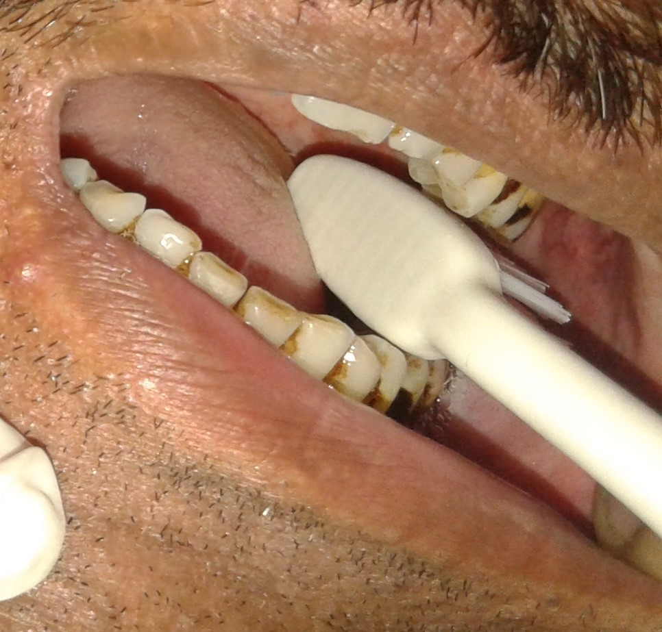
Figure 1: Oral brush cytology in a patient with benign oral lesion (using a tooth brush).
The accuracy, sensitivity and specificity of oral brush cytology for malignant tumors were calculated and found to be 86.36%, 81.25% and 100% respectively. Whereas brush cytology for detection of benign and inflammatory conditions revealed that all the 15 cases of benign tumors and 06 cases as inflammatory were confirmed as benign and inflammatory on biopsy respectively without any variation.
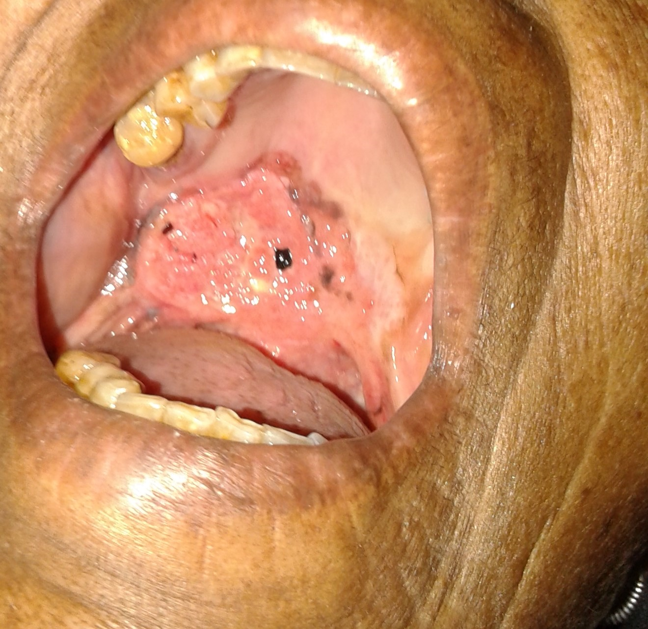
Figure 2: Gross appearance of an oral malignant lesion.
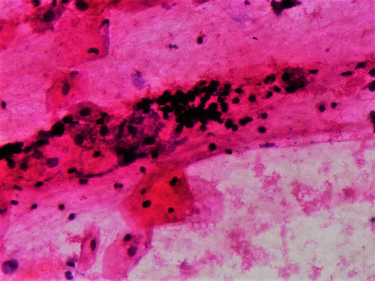
Figure 3: Oral brush cytology smear showing malignant cell.

Figure 4: Histological section showing well-differentiated squamous cell carcinoma (H&E; 40X)
Squamous cell carcinoma of the oral cavity constitutes 90% of all oral cancers.1 The incidence of oral cancer is 2% to 4% in the western countries while it is very high in south Asian countries like Pakistan and India. In India, OSCC accounts for 40% of all cancers,14 while in Pakistan the prevalence of oral cancer is highest than rest of the world.15
The diagnostic accuracy of the brush biopsies was calculated in a study by Remmerbach et al16 reporting a sensitivity of 97.8% and specificity of 100%. Another study by Maraki and colleagues17 exhibited cytology along with DNA cytometry to be highly specific, sensitive and non-invasive technique resulting in 100% sensitivity. An additional study also established a high sensitivity (92.5%) and specificity (100%) of the cytological diagnosis for OSCC. Driemel et al18 estimated the oral brush biopsies performance by means of standard morphological analysis to detect OSCC and their respective precursor lesions, with a sensitivity of 79% and specificity of 93% respectively. In another study of Remmerbach and coworkers19 a high diagnostic accuracy of oral brush cytology in doubtful oral lesions was reported with a specificity of 99.5% and sensitivity of 94.6%. Babshet et al20 stated that the oral brush cytology had a sensitivity of 71.4%, using oral CDx technique. A UK audit with hindsight established the specificity, sensitivity and negative and positive predictive rates of brush cytology in the diagnosis of pre-malignant lesions in 112 cases exhibited a sensitivity of 71.4% in detection of OSCC or dysplasia. Present study is consistent with all these studies, as it shows an accuracy of 86.3%, sensitivity of 81.25% and specificity of 100%.
Thus, oral brush cytology followed by biopsy and histopathological examination have a significant role in diagnosis and can be recommended for all patients exposed to risk factors of developing oral cancer, for example tobacco chewers. Oral brush cytology may as well be applied as a population screening test for early detection of squamous cell carcinoma of oral cavity. It can also help in follow up after operation. In this way oral brush cytology could help in improving the five-year survival rate.
The present study concludes that oral brush cytology has good accuracy, sensitivity and specificity for detection of oral epithelial lesions and can be useful in early detection of oral cancer as well as other lesions. Brush cytology is an easy and painless procedure, and due to its non-invasiveness can be a good screening method for detection of oral lesions.
- Haines, GK "Pathology of Head and Neck Cancers I: Epithelial and Related Tumors". Head & Neck Cancer: Current Perspectives, Advances, and Challenges. Springer Science & Business Media. 2013;257–87. Doi: 10.1007/978-94-007-5827-8
- Chi AC, Day TA, Neville BW. Oral cavity and oropharyngeal squamous cell carcinoma—an update.CA cancer J Clin 2015; 65(5):401–21. Doi: 10.3322/caac.21293
- Porcheri C, Meisel CT, Mitsiadis T. Multifactorial Contribution of Notch Signaling in Head and Neck Squamous Cell Carcinoma. Int. J. Mol. Sci. 2019, 20(6), 1520. Doi: 10.3390/ijms20061520
- Naghavi M, Wang H, Lozano R, Davis A, Liang X, Zhou M, et al. Global, regional, and national age–sex specific all-cause and cause-specific mortality for 240 causes of death, 1990–2013: a systematic analysis for the Global Burden of Disease Study 2013. Lancet 2015; 385(9963): 117–71. Doi: 10.1016/S0140-6736(14)61682-2
- Martin-Hernan F, Sanchez-Hernandez JG; Cano J, Campo J, Del Romero J. "Oral cancer, HPV infection and evidence of sexual transmission". Med Oral, Patol Oral Cir Bucal 2013;18(3): 439–44. Doi: 10.4317/medoral.18419
- Cancer Institute. SEER Stat Fact Sheets: Oral Cavity and Pharynx Cancer. Available at: http://seer.cancer.gov/statfacts/html/oralcav.html. 2015
- Maher NG, Collgros H, Uribe P, Ch’ng S, Rajadhyaksha M, Guitera P. In vivo confocal microscopy for the oral cavity: Current state of the field and future potential. Oral Oncol, 2016, 54 28-35. Doi: 10.1016/j.oraloncology.2016.01.003
- Fang QG, ShiS, Li ZN, Zhang X, Liua FY, Xu ZF et al. Squamous cell carcinoma of the buccal mucosa: Analysis of clinical presentation, outcome and prognostic factors. Mol Clin Oncol. 2013; 1(3): 531-34. Doi: 10.3892/mco.2013.86
- Keshavarzi M, Darijani M, Fatemeh M. Molecular Imaging and Oral Cancer Diagnosis and Therapy. J. Cell Bioche. 2017; 118(10): 3055-60. Doi: 10.1002/jcb.26042
- Kumaresan GD, Jagannathan N. Exfoliative cytology-A predictive diagnostic tool. Int J Pharma Pharm Sci. 2014;6(5):1-3
- Chaturvedi P, DattaS, Arya S, Rangarajan V, Kane SV, NairD et.al. Prospective study of ultrasound‐guided fine‐needle aspiration cytology and sentinel node biopsy in the staging of clinically negative T1 and T2 oral cancer. Head Neck-J Sci Spec. 2015; 37(10):1504-8. Doi: 10.1002/hed.23787
- Yamane, Taro. Statistics: An Introductory Analysis, 2nd Edition, New York: Harper and Row. 1967
- Baratloo A, Hosseini M, Negida A, El Ashal G. Part 1: Simple Definition and Calculation of Accuracy, Sensitivity and Specificity. Emerg(Tehran)., 2015; 3(2): 48-9. PMID: 26495380
- Mehrotra R, Singh M, Kumar D, Pande AN, Gupta RK, Sinha US. Age specific incidence rate and pathological spectrum of oral cancer in Allahabad. Ind J Med Sci. 2003;57(9):400–4. PMID: 14515030
- Siddiqui PQR. Oral Cancer: A New Epidemic Can Be Foreseen in Pakistan, Pakistan Journal of Medicine and Dentistry 2013, 2 (01): 1-2
- Remmerbach TW, Mathes SN, Weidenbach H, Hemprich A, Bocking A. Noninvasive brush biopsy as an innovative tool for early detection of oral carcinomas. Mund Kiefer Gesichtschir. 2004; 8:229-36. Doi: 10.1007/s10006-004-0542-z
- MarakiD, Becker J, Boecking A. Cytologic and DNA-cytometric very early diagnosis of oral cancer. J Oral Pathol Med 2004; 33(7):398-404. Doi: 10.1111/j.1600-0714.2004. 0235.x
- Driemel O, Kunkel M, Hullmann M, Kleinsasser N, Staudenmaier R, Muller-Richter U, et al. Performance of conventional oral brush biopsies. HNO. 2008; 56(2):205-10. Doi: 10.1007/s00106-007-1662-1
- Remmerbach TW, Weidenbach H, Muller C, Hemprich A, Pomjanski N, Buckstegge B et al. Diagnostic value of nucleolar organizer regions (AgNORs) in brush biopsies of suspicious lesions of the oral cavity. Anal Cell Pathol. 2003;25(3):139-46. PMID: 12775918
- Babshet M, Nandimath K, Pervatikar SK, NaikmasurVG. Efficacy of oral brush cytology in the evaluation of the oral premalignant and malignant lesions. J Cytol. 2011; 28(4): 165–172. Doi: 10.4103/0970-9371.86342