Immunohistochemical Expression of BCL-2 in Malignant Salivary Gland Tumors
Faiz Rasul1, Sultan Muhammad Wahid2, Iman Imran3, Zainab Rizvi4,Rozina Jaffar5,Ambereen Anwar6,
1 Assistant Professor, Department of Oral Pathology, de’Montmorency College of Dentistry, Lahore Pakistan
2 Head, Department of Dentistry, Lahore General Hospital, Lahore Pakistan
3 Final Year MBBS, CMH Lahore Medical College, Lahore Pakistan
4 Associate Professor, Department of Oral Pathology, de’Montmorency College of Dentistry, Lahore Pakistan
5 Professor, Department of Pathology, Rahbar Medical & Dental College, Lahore Pakistan
6 Professor, Department of Pathology, Allama Iqbal Medical College, Lahore Pakistan
Background:
Malignant salivary gland tumors (MSGTs) consist of a heterogeneous group of neoplasms with complex clinicopathological features and biological behaviors. The purpose of this study was to determine the expression of Bcl-2 antiapoptotic protein in mucoepidermoid carcinoma (MEC), adenoid cystic carcinoma (ADCC), acinic cell carcinoma (ACC) and polymorphous low-grade adenocarcinoma (PLGA) of salivary glands and to find out its association with different grades of these tumors.
Material and Methods: This descriptive study included 55 cases of MSGTs. Tissue sections were stained with routine hematoxylin and eosin stain as well as Bcl-2 immunostain. MSGTs were graded as low grade (Low grade MEC, ACC, PLGA, and tubular pattern of ADCC), intermediate grade (cribriform pattern of ADCC, and intermediate grade of MEC) and high grade (high grade of MEC and solid pattern of ADCC) tumors on H&E sections. Bcl-2 expression was scored as ‘negative’ (<5% of neoplastic cells), ‘1’ (5-19% of neoplastic cells), ‘2’ (20-49% of neoplastic cells), and ‘3’ (>50% of neoplastic cells), respectively.
Results: : MSGTs most commonly involved the parotid gland (52.7%), while ADCC (40%) and MEC (38.2%) were the most common tumors. Expression of Bcl-2 was strongly positive in 56.4% cases of MSGTs which included ADCC (71%), MEC (19.4%) and ACC (9.7%), respectively. A significant association was found between Bcl-2 staining and types of MSGTs i.e., MEC, ADCC, ACC (P = .001) as well as between Bcl-2 staining and grades of MSGTs (P = .013).
Conclusions:Bcl-2 protein is expressed in malignant salivary gland tumors. Its expression maybe helpful in grading small biopsies, predicting behavior, and planning targeted therapy of MSGTs.
Key words: Bcl-2, Immunohistochemistry, Malignant salivary gland tumors.
Salivary glands tumors (SGTs) are rare tumors and their distribution varies according to geographic location and race1, 2. These comprise approximately 3-10% of the neoplasms of the head and neck region and 1 % of the neoplasms of the whole body3. Annual global incidence of SGTs is between 0.4 to 13.5 cases per 100 000 individuals. Among these, malignant salivary gland tumors (MSGTs) are 0.40 to 0.65 per 100 000 cases4. MSGTs are 0.3% of all malignancies5. Early diagnosis of MSGTs plays an important role in better management of these tumors6. Different diagnostic procedure have been used such as diffusion-weighed magnetic resonance imaging and fluorodeoxyglucose positron emission tomography/CT which show good diagnostic accuracy in identifying local recurrence and distant metastases in MSGTs7. Fine-needle aspiration cytology (FNAC) is also used as a screening or early diagnostic tool in these tumors, however its role is limited3. Core needle biopsy and intraoperative frozen section are also helpful techniques.
Accurate diagnosis depends upon the histological evaluation of MSGTs. The histopathological diagnosis of these tumors is usually made through the assessment of histological architecture, cellular features and differentiation, component of tumor stroma, the growth pattern of the tumor borders, along with the clinical information9. MSGTs are comprised of a heterogeneous population of cells and show different architectural patterns which makes them complex histopathologically10 rendering diagnosis difficult even by expert histopathologists. Although hematoxylin-eosin (H&E) staining is still the gold standard for diagnosing the MSGTs, yet its role is limited in certain cases. Immunohistochemistry (IHC) can enhance the accuracy and may be a helpful tool when there is a need to investigate cell type and differentiation status, cell proliferation, and tumor protein expression that cannot be assessed by routine histological examination alone. There is also a need to investigate new biomarkers that can be useful in the diagnosis and grading of malignant salivary gland tumors11.
Bcl-2 (the B-cell lymphoma) oncoprotein is a useful marker for investigation in MSGTs. Apoptosis is a genetically predetermined process which is involved in the deletion of cells in normal tissues as well as in malignant tumors. It may be elicited by several molecular pathways. BCL-2 gene family consists of different regulators involved in apoptosis. These proteins are divided according to their function into two groups, namely the anti- and pro-apoptotic proteins. The Bcl-2, Bcl-xL and Mcl-1 are considered as anti-apoptotic proteins, while Bax, Bak, Bad and Bcl-xS promote apoptosis and are considered as pro-apoptotic proteins12. Deregulation of these genes controlling apoptosis may contribute to the process of tumor genesis by reducing the rate of cell death and allowing the accumulation of other genetic defects. Frequently apoptosis is dysregulated in human cancers, which makes it a suitable target for anticancer therapy. Bcl-2 proteins play a key role in preventing programmed cell death by favoring prolonged survival in normal and neoplastic cells. Its increased expression has been seen in a number of tumors and is related to resistance to conventional cancer management13. The present study was planned to determine the expression of Bcl-2 proteins in MEC, ADCC, ACC, and PLGA of salivary gland and to determine association of BCL-2 expression with different grades of these tumors.
After the approval of research project from the ethical board of Postgraduate Medical Institute, Lahore and University of Health Sciences Lahore, Pakistan, this descriptive study was carried out on diagnosed cases of MSGTs (n=55) during a two years period (January 2015 to December 2016). Samples were collected from the Department of Surgery Lahore General Hospital, Lahore, Oral and Maxillofacial department of Mayo Hospital, Lahore and de’Montmorency College of Dentistry, Lahore through non probability purposive sampling, after taking informed consent from the patients. Recurrent cases and patients on radiotherapy or chemotherapy were excluded from this study. Clinical data such as age, gender and demographic characteristics of each individual along with site and size of MSGTs were also recorded in a proforma. Standard procedure of gross examination of specimen size, color and consistency were recorded as per protocol. Routine H&E and immunostains were performed. Two histopathologists examined MSGT for their histological type, grade, lymphovascular invasion and margin of tumor on Hematoxylin and eosin (H&E) slides.
Grading of tumor was done as low grade (LG), intermediate (IG) and high grade (HG) tumors. LG tumors included low grade MEC, ACC, PLGA, and tubular pattern of ADCC. IG tumors included cribriform pattern of ADCC, and intermediate grade of MEC while HG tumors included high grade of MEC and solid pattern of ADCC5.
The expression of Bcl-2 was evaluated immunohistochemically. Bcl-2 immunoreactivity was divided into four groups as follows:14 Score ‘0’ or negative had less than 5% stained neoplastic cells, score ‘1’ or weak positive (WP) had 5-19% stained neoplastic cells, score ‘2’ or moderate positive (MP) had 20-50% neoplastic cells stained while score ‘3’ or strong positive (SP) had more than 50% neoplastic cells stained. Observations were made on the basis of intensity of cytoplasmic staining. Data were entered and analyzed by using SPSS version 21. Chi-square test was used to determine the association between Bcl-2 expression across different types and grades. P-value ≤ .05 was considered as statistically significant.
Results showed that MSGTs were seen more frequently in females, with a male-to-female ratio of 5:6. The mean age of the patients was 50 ± 4.03 years. Parotid gland was the most commonly involved gland (52.7%), followed by minor salivary glands (29.1%). The data showed no major difference between right and left sides. ADCC (40%) and MEC (38.2%) were the most common tumors followed by ACC (12.7%) and PLGA (9.1%), respectively.
Details of comparison of BCL-2 staining with tumor types and tumor grades are shown in Tables II. A statistically significant association was found between BCL-2 staining and types of carcinomas (P < .001). A statistically significant association was found between Bcl-2 expression and grades of tumor (P = .013). Most of the low-grade tumors (75%) had weak Bcl-2 positivity, intermediate grade had moderate (33.3%), while high grade tumors had mostly strong Bcl-2 positivity (Table II; Figures 1 & 2).
Manjunatha et al., reported 78% Bcl-2 expression in MSGTs with 100% expression in ADCC, similar to current study. They concluded that on varying intensity of Bcl-2 there is not much difference in ADCC with respect to grading and type. Whereas, in the current study, contrary to results above a significantly high association was found in Bcl-2 staining and types of carcinomas as well as grade of cancer 15.
Kaur and Gupta reported weak to strong positive Bcl-2 expression in MSGTs. Bcl-2 expression in MEC was weakly positive in 4 cases and strong positive in one case. However, in the current study, total cases of MEC were 21, out of them 7 expressed weak positive staining, 8 expressed moderate staining and 6 expressed strong positive staining. PLGA had weak positive expression of Bcl-2 in both the studies16. Al-Rawi et al. described positive expression of Bcl-2 in all cases of MSGTs, with high-grade Adenocystic carcinomas showing highest Bcl-2 expression (90%). Its expression was lowest in low-grade cystic MEC17.
Table I: Descriptive Statistics of Malignant Salivary Gland Tumors (n=55) |
||
Variables |
Subgroups |
n (%) |
Age |
20- 40 |
19 (34.5%) |
41-60 |
17 (30.9%) |
|
61-80 |
19 (34.5) |
|
Gender |
Male |
25 (45.5) |
Female |
30 (54.5) |
|
Occupation |
Industry |
10 (18.2) |
Farmer |
8 (14.5) |
|
Labor |
14 (25.5) |
|
Office job |
1 (1.8) |
|
Household |
22 (40) |
|
Hospital |
Mayo hospital |
17 (30.9) |
Lahore General Hospital |
20(36.4) |
|
de'Montmorency College of Dentistry/ PDH |
18(32.7) |
|
Site |
Parotid Gland |
29 (52.7) |
|
Submandibular salivary Gland |
9 (16.4) |
Sublingual salivary Gland |
1 (1.8) |
|
Minor salivary gland on palate |
10 (18.2) |
|
Minor salivary gland on tongue |
1 (1.8) |
|
Minor salivary gland on labial mucosa |
1(1.8) |
|
Minor salivary gland on Buccal mucosa |
4 (7.3) |
|
Laterality |
Right |
28 (50.9) |
Left |
27 (49.1) |
|
Specimen Type |
Incisional |
10 (18.2) |
Excisional |
40 (72.7) |
|
Resection |
5 (9.1) |
|
Size |
Less than 1cm maximum diameter |
2 (3.6) |
1cm to 2cm maximum diameter |
4 (7.3) |
|
2-5cm |
42 (76.4) |
|
More than 5 cm in maximum diameter |
7(12.7) |
|
Mass |
Solid |
55 |
Type of Tumor |
Mucoepidermoid Carcinoma |
21 (38.2) |
Adenoid Cystic Carcinoma |
22 (40.0) |
|
Acinic Cell Carcinoma |
7 (12.7) |
|
Polymorphous Low-Grade Adenocarcinoma |
5 (9.1) |
|
Grade |
Low Grade |
22 (40.0) |
Intermediate |
13 (23.6) |
|
High Grade |
20 (36.4) |
|
Invasion |
No lymphovascular or neural invasion |
31 (56.4) |
Neural invasion |
22 (40.0) |
|
Both lymphovascular or neural invasion |
2(3.6) |
|
Bcl-2 Expression |
+ weak positive [staining in 5-19% of neoplastic cells] |
12 (21.8) |
++ moderate positive [staining in 20-50% of neoplastic cells] |
12 (21.8) |
|
+++ strong positive [staining in more than 50% of neoplastic cells] |
31 (56.4) |
|
Table II: Comparison of Bcl-2 staining with types and grades of MSGTs |
||||
Type of Tumor |
Bcl-2 Positivity |
Total |
||
Weak |
Moderate |
Strong |
||
MEC |
7 (58.3%) |
8 (66.7%) |
6 (19.3%) |
21 (38.2%) |
ACC |
0 (0%) |
4(33.3%) |
3 (9.7%) |
7 (12.7%) |
ADCC |
0 (0%) |
0 (0%) |
22 (71%) |
22 (40%) |
PLGA |
5 (41.7%) |
0 (0%) |
0 (0%) |
5 (9.1%) |
Total |
12 |
12 |
31 |
55 |
Tumor Grade |
Bcl-2 Positivity |
|
||
Weak |
Moderate |
Strong |
Total |
|
Low |
9 (75%) |
6 (50%) |
7 (22.6%) |
22 (40%) |
Intermediate |
1 (8.3%) |
4 (33.3%) |
8 (25.8%) |
13 (23.6%) |
High |
2 (16.7%) |
2 (16.7%) |
16 (51.6%) |
20(36.4%) |
Total |
12 |
12 |
31 |
55 |
However, in the current study, almost all cases of MEC (n=21) had Bcl-2 expression. In a study conducted by Soini et al. expression of Bcl-2 was strong positive in ADCC as compared to MEC and ACC. In our study all cases of ADCC showed strong positive expression of Bcl-2. Similarly, we also observed BCL-2 expression in all cases of MSGTs in contrast to Soini et al18. Carlinfante et al. reported a high expression of Bcl-2 (almost 90%) in ADCC, which is similar to current study19.
We found a statistically significant association between Bcl-2 expression and MSGTs. Furthermore, significant association was also found between expression of Bcl-2 and grades of MSGTs. Hellquist et al. did not report any statistically significant difference between expression of Bcl-2 and TUNEL in ACC. They concluded that expression of Bcl-2, MIB-1, clinical staging and TUNEL are helpful prognostic tools for predicting prognosis of acinic cell carcinoma20. On the other hand, in our study Bcl-2 was statistically significant with type and grades of MSGTs.
Janjua et al. reported positive expression of Bcl-2 in 60% of MEC and expression of BCL-2 was strong positive in low grades and weak positive for high grades of MEC. However, in our study, high grade MEC expressed strong positivity in most of the cases. Out of 10 high grades MEC, 6 were strong positive, 2 were intermediate positive and 2 were weak positive. While a significant association was seen between expression of Bcl-2 and grades of MEC in both the studies21. Jiang et al. aimed to determine the expression of Bcl-2 in ADCC (n=35) and reported its expression in 60% of tumors. Although the sample size of ADCC in the current study was smaller (n=22) but all cases of ADCC expressed Bcl-2 positivity as strong positive. In the current study a statistically significant association was found between Bcl-2 positivity, type, and grades of MSGTs in contrast to study by Jiang et al22.
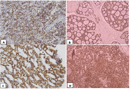
Figure 1: Photomicrographs of Bcl-2 Immunostaining of Malignant Salivary Glands Tumors. A shows high grade Mucoepidermoid carcinoma with strong positive expression (x 400). B shows intermediate grade Adenoid cystic carcinoma with strong positive expression at low power (x 100) and C shows strong Bcl-2 positivity at high power (x 400). D shows BCL-2 immunostaining of low grade Acinic cell carcinoma with strong positive expression (x 100)
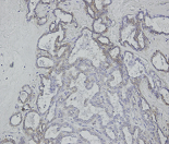
Figure 2: Bcl-2 immunostaining of Polymorphous low-grade adenocarcinoma showing weak positive expression (x 200).
Norberg-Spaak et al. determined the biological behavior of ADCC in its three subtypes; solid, cribriform, and tubular by using Bcl-2 and its expression was statistically insignificant with grades of tumors which is in contrast to the current study23. Yin et al. analyzed Bcl-2 expression in 71 cases of MEC of minor salivary glands. They reported that low grade MEC had higher expression of Bcl-2 as compared to intermediate and high grade MEC. This study reveals that higher the grade of MEC, stronger is the expression of Bcl-2 in contrast to Yin’s findings24. Atarbashi reported 30% expression of Bcl-2 in MEC which is lower than this study25.
Rasul et al. conducted a study on 35 cases of ADCC in Lahore, Pakistan and reported that these tumors were more common in females mostly in the fourth to sixth decade26. In major salivary glands, parotid gland was the most common site while in minor salivary glands the most common site was palate. Majority cases reported as excisional biopsies (54.3%) with sizes ranging between 2 cm to 5 cm (68.8%). Histopathologically, 19 cases (54%) were categorized as high-grade tumors. All cases showed expression of Bcl-2 irrespective of the grade of the tumor. Expression of Bcl-2 was strongly positive in all cases of ADCC26.
Epidemiological features of MSGTS have also been reported locally in Pakistan by different authors. A study conducted in Jamshoro Pakistan reported that mucoepidermoid carcinoma was the most common malignant salivary gland tumor2. In the current study, ADCC was more common than MEC of the salivary glands’ tumors. Recently a study was conducted in Armed Forces Institute of Pathology on 30 cases of MEC and it was reported that low grade MEC was most common tumor similar to result of this study28.
There was certain limitation of the current study, which might be the cause of differences in results while comparing with other studies. Malignant salivary gland tumors are rare, therefore the sample size in the current study was small (n=55) so, it was difficult to evaluate the definitive role of Bcl-2. Similarly, there was an unequal distribution of the different types and grades of these tumors. The distribution of these tumors was also unequal in the minor and major salivary glands. Further studies with larger sample size are recommended to find out the definitive role of Bcl-2 in benign and malignant salivary gland tumors.
Most of the low-grade tumors express weak positivity of Bcl-2 while most of the high-grade tumors express strong positivity in malignant salivary gland tumors. Positive expression of Bcl-2 in malignant salivary gland tumors can help in predicting the behavior of these tumors regarding their potential for aggressiveness. In addition, molecular targeted therapy against BCL-2 can be planned in future for better management of salivary gland tumors.
We are thankful to Mr. Amin and Mr. Sohail for their technical help in slide preparation for Bcl-2 immunostaining.
- Araya J, Martinez R, Niklander S, Marshall M, Esguep A. Incidence and prevalence of salivary gland tumours in Valparaiso, Chile. Med Oral Patol Oral Cir Bucal. 2015; 20(5): 532-9. Doi: 10.4317/medoral.20337.
- Sando Z, Fokouo JV, Mebada AO, Djomou F, NDjolo A, Oyono JLE. Epidemiological and histopathological patterns of salivary gland tumors in Cameroon. Pan Afr Med J. 2016; 23(1): 1-66. Doi: 10.11604/pamj.2016.23.66.5105.
- Carlson ER, Schlieve T. Salivary gland malignancies. Oral and Maxillofacial Surgery Clinics. 2019; 31(1): 125-44. Doi: 10.1016/j.coms.2018.08.007.
- Faur, AC, Sas I, Motoc AGM, Cornianu M, Zamfir CL, Lazar DC, et al. Ki-67 and p53 immunostaining assessment of proliferativeactivity in salivary tumors. Rom J Morphol Embryol. 2015; 56(4): 1429–39. PMID: 26743291.
- Stenner M, Klussmann JP. Current update on established and novel biomarkers in salivary gland carcinoma pathology and the molecular pathways involved. Eur Arch Otorhinolaryngol. 2009; 266(3): 333–41. Doi: 10.1007/s00405-008-0882-7.
- Gupta A, Koochakzadeh S, Neskey DM, Nguyen SA, Lentsch EJ. Salivary Carcinosarcoma: An Extremely Rare and Highly Aggressive Malignancy. The Laryngoscope. 2020; 130(5): E335-9. Doi: 10.1002/lary.28183.
- Thakkar M, Brahmbhatt P, Raychaudhuri C. Evaluation of salivary gland tumors by ultrasonography and CT scan. IAIM. 2017; 4(4): 14-18.
- Oroz A, Bokun Z, Antonijević Đ, Jevđić J. The determination of specificity, sensitivity and accuracy of core needle biopsy in the diagnosis of parotid and submandibular salivary glands tumors. Vojnosanitetski pregled. 2019; 76(9): 921-8. Doi: 10.2298/VSP170320001O.
- Nagao T, Sato E, Inoue R, Oshiro H, Takahash RH, Nagai T et al. Immunohistochemical Analysis of Salivary Gland Tumors: Application for Surgical Pathology Practice. Acta Histochem. Cytochem. 2012; 45(5): 269–82. Doi: 10.1267/ahc.12019.
- Moutasim KA, Thomas GJ. Salivary gland tumours: update on molecular diagnostics. Diagnostic Histopathology. 2020; 26(4). Doi: 10.1016/j.mpdhp.2020.01.002.
- Ibrahim TR, Ahmed MM, Hegazy AA. Diagnostic Utility of Immunohistochemical Expressions of IMP3 Versus DOG1 and p63 in Salivary Gland Tumors. Turk Patoloji Derg. 2020; 36(3). 227-236. doi: 10.5146/tjpath.2020.01496.
- Xie X, Nordgård S, Clausen F, Petter O, Boysen M. Prognostic Significance of Bax and Bcl-2 Expressions in Adenoid Cystic Carcinoma of Major and Minor Salivary Glands of Nasal and Oral Epithelium. Open Otorhinolaryngol. J. 2010; 4(1): 20-26. Doi: 10.2174/1874428101004010020.
- Abdulhussain MM, Mohsen AS. Immunohistochemical Evaluation of Bcl-2 in Mucoepidermoid Carcinoma and Adenoid Cystic Carcinoma of Salivary Glands. Journal of International Dental and Medical Research. 2020; 13(1): 180-7.
- AL-Rawi NH, Ahmed JN, Mohammed HO. Immunohistochemical Expression of P53 and Bcl-2 in benign and malignant salivary glands tumors. MDJ. 2007; 4 (1): 1-10.
- Manjunatha B, Kumar G, Raghunath V. Immunohistochemical expression of Bcl-2 in benign and malignant salivary gland tumors. Med. Oral Patol. Oral Cir. Bucal. 2011; 16(4): 503-7. Doi: 10.4317/medoral.16.e503.
- Kaur H, Gupta S. An Analysis of the Expression of Bcl-2, Podoplanin and Lymph Angiogenesis in Benign and Malignant Salivary Gland Tumours. J Clin Exp Pathol. 2013; 3(3): 145. Doi: 10.4172/2161-0681.1000145.
- AL-Rawi NH, Omer H, Al Kawas S. Immunohistochemical Expression of P53 and Bcl-2 in benign and malignant salivary glands tumors. J Oral Pathol Med. 2010; 39(1): 48-55. Doi: 10.1111/j.1600-0714.2009.00816.x.
- Soini Y, Tormanen U, Paakko P. Apoptosis is inversely related to Bcl-2 but not to Bax expression in salivary gland tumors. Histopathology. 1998; 32(1): 28-34. Doi: 10.1046/j.1365-2559.1998.00334.x.
- Carlinfante G, Lazzaretti M, Ferrari S, Bianchi B, Crafa P. P53, Bcl-2 and Ki-67 expression in adenoid cystic carcinoma of the palate. A clinico-pathologic study of 21 cases with long-term follow-up. Pathol Res Pract. 2005; 200(11-12): 791-9. Doi: 10.1016/j.prp.2004.08.008.
- Hellquist HB, Sundelin K, Di Bacco A, Tytor M, Manzotti M, Viale G. Tumour growth fraction and apoptosis in salivary gland acinic cell carcinomas. Prognostic implications of Ki-67 and Bcl-2 expression and of in situ end labelling (TUNEL). J. Pathol. 2008; 215(4): 458. Doi: 10.1002/(SICI)1096-9896(199703)181:3<323::AI D-PATH780>3.0.CO;2-K.
- Janjua OS, Qureshi SM, Khan TS, Alamgir W. Bcl-2 protein expression in mucoepidermoid carcinoma of salivary glands: a single institution experience. Hematol Oncol Stem Cell Ther. 2012 5(2): 96-100. Doi: 10.5144/1658-3876.2012.96.
- Jiang LC, Huang SY, Zhang DS, Zhang SH, Li WG, Zheng PH, Chen ZW. Expression of beclin 1 in primary salivary adenoid cystic carcinoma and its relation to Bcl-2 and p53 and prognosis. Braz. J Med Biol Res. 2014; 47(3): 1-6. Doi: 10.1590/1414-431X20133231.
- Norberg-Spaak L, Dardick I, Ledin T. Adenoid cystic carcinoma: use of cell proliferation, BCL-2 expression, histologic grade, and clinical stage as predictors of clinical outcome. Head Neck. 2000; 22(5): 489–97. Doi: 10.1002/1097-0347(200008)22:5<489::aid-hed8>3.0.co;2-x.
- Yin HF, Okada N, Takagi M. Apoptosis and apoptotic-related factors in mucoepidermoid carcinoma of the oral minor salivary glands. Pathol Int. 2001; 50(8): 603-9. Doi: 10.1046/j.1440-1827.2000.01091.x.
- Atarbashi S, Elahi M, Khani M, Rakhshan V. Immunohistochemical analysis of B-cell lymphoma -2 in pleomorphic adenoma and mucoepidermoid carcinoma. Dent Res J. (Isfahan). 2014; 11(2): 257–63. PMID: 24932199.
- Rasul F, Rizvi Z, Wahid SM, Haseeb MT, Jaffar R, Amjad A. Immunohistochemical Expression of BCL-2 in Adenoid Cystic Carcinoma of Salivary Gland Tumors. Med Forum. 2018; 29(12): 14-19.
- Baloch MA, Ujjan I, Munawar M. Demography and Histopathological presentations of oral and maxillo-facial region tumours in Jamshoro, Pakistan. J Muhammad Med Coll. 2018; 9(2): 81-4.
- Niazi Z, Khan I, Farhan F, Ilyas M, Ali M, Hasan SR. Mucoepidermoid Carcinoma: A Demographic and Histopathological Profile. J Islamabad Med Dental Coll. 2020; 9(4): 298-302. Doi: 10.35787/jimdc.v9i4.460.

An Official Publication of
Islamabad Medical & Dental College
Volume 10 Issue 1
Author's Contribution:
1Conception; Literature research; manuscript design and drafting; 2,3 Critical analysis and manuscript review; Data analysis; Manuscript Editing.
Article info:
Received: April 27, 2020
Accepted: March 18, 2021
Funding Source: Nil
Conflict of Interest: Nil
Address of Correspondence
Faiz Rasul
Email:
bayfaiz871@gmail.com
Cite this article. Rasul F, Wahid SM, Imran I, Rizvi Z, Jaffar R, Anwar A. Immunohistochemical Expression of BCL-2 in Malignant Salivary Gland Tumors. J Islamabad Med Dental Coll. 2021; 10(1): 15-22. Doi: 10.35787/jimdc.v10i1.542