Histopathology Images
Melanoma |
|
 |
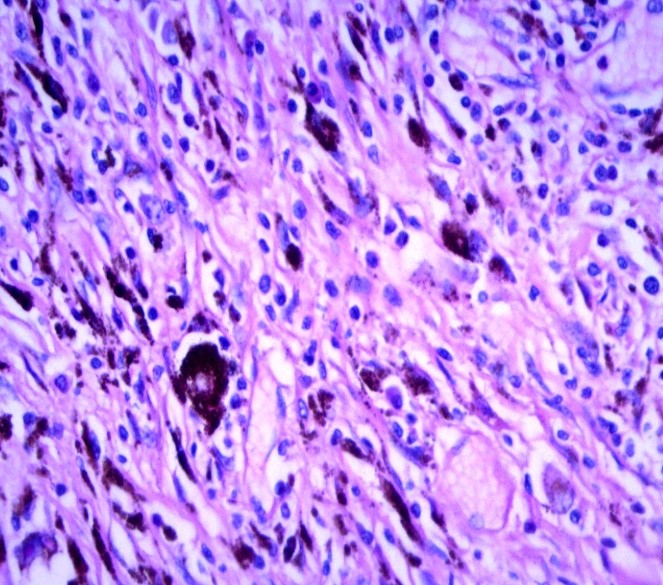 |
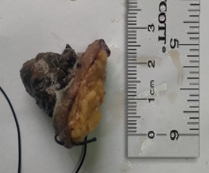
Figure 1: Gross appearance of Melanoma. Skin with large protruded pigmented lesion measuring approximately 2cm in high. |
Figure 2: Microscopic view of melanoma cell nest with abundant melanin. The cells appear markedly pleomorphic (H&E 400). |
 |
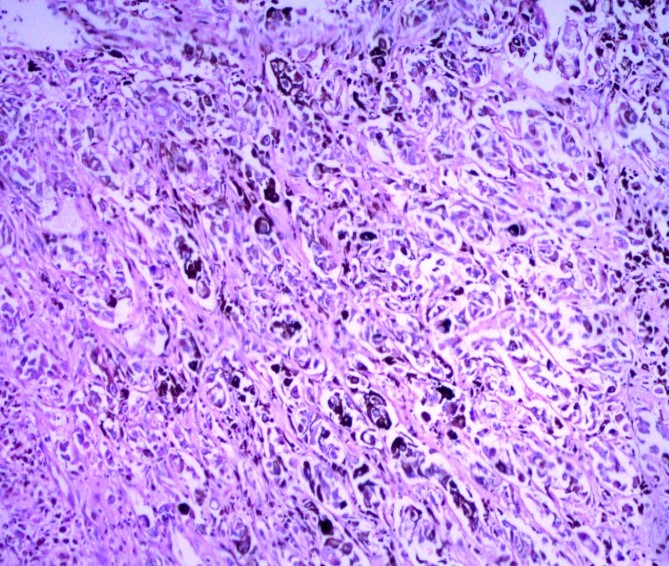 |
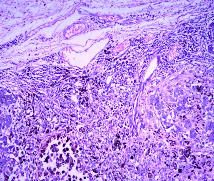
| Figure 3: Nest of Melanotic cells with melanin. | Figure 4: High power microscopic view showing nest of melanotic cells with blackish melanin pigmentation. |
Polypoidal Leiomyoma |
|
 |
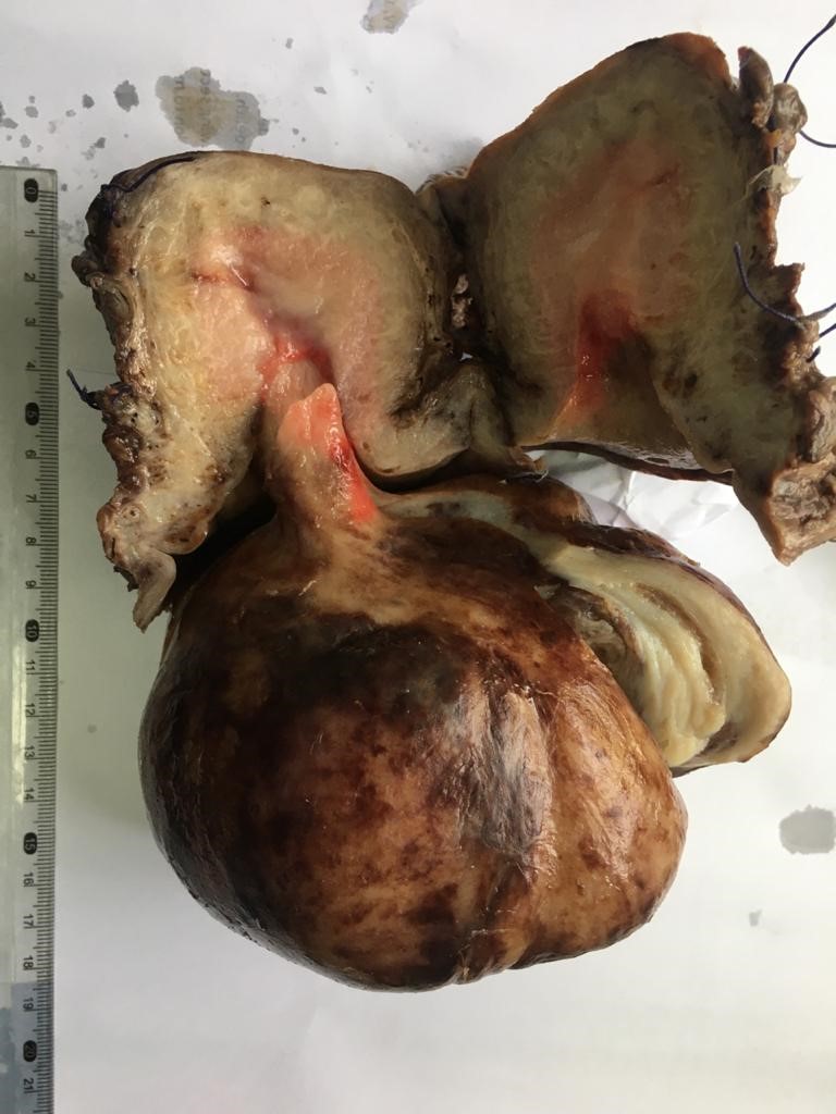 |
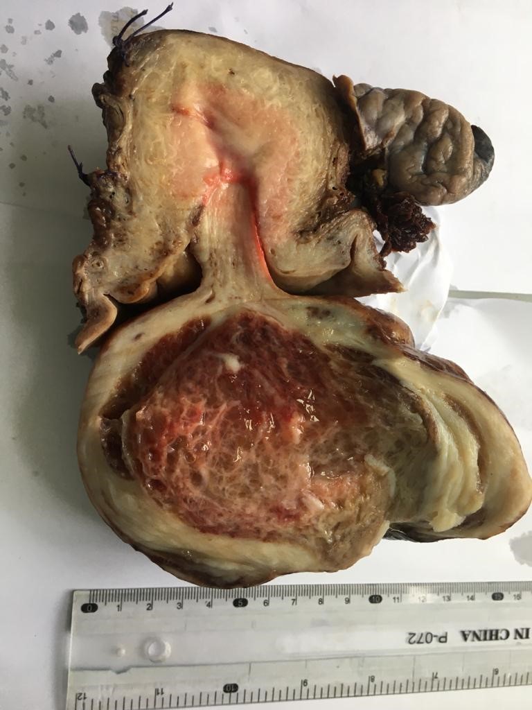
| Figure 1: Cut surface of leiomyoma with myxoid degeneration. | Figure 2: Large polypoid leiomyoma hanging from the cervical cavity. |
 |
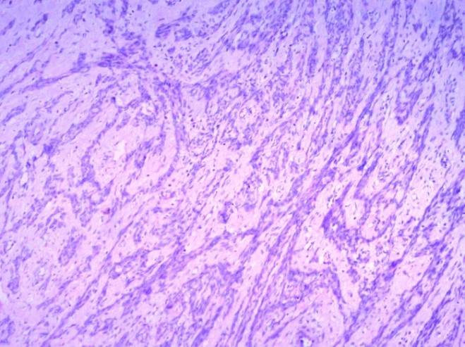 |
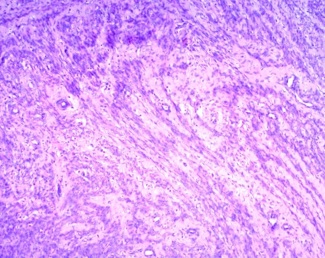
| Figure 3: Microscopic view. Fascicles of smooth muscles cells with myxoid stroma (H&E 100). | Figure 4: Fascicles of smooth muscles with bland looking cells and myxoid areas (H&E 100). |

An official publication of
Islamabad Medical & Dental College
Volume 10 Issue 1
Contributed by:
Prof Dr. Saeed Alam, Dr. Javeria Faridi
Department of Pathology