Comparative Benefits of Tissue Marking by Poster Ink in Histopathology
Ayesha Sarwar 1 , Ashok Kumar Tanwani 2 , Anum Usman 3 , Khalida Moeed 4 .
1Assistant Professor, Department of Pathology, HBS Medical and Dental College, Islamabad.
2 HOD Pathology, HBS Medical and Dental College, Islamabad.
3Associate Professor, Department of Pathology, Al Nafees Medical College, Islamabad.
4Assistant Professor, Department of Anatomy, Loralai Medical College, Balochistan
Background:Tissue margin marking with India ink is important in decision making for surgeons. The present study was
conducted to examine the reliability of different shades of locally available poster colours in tissue marking and to
evaluate the colour perceptibility microscopically in comparison with similar tissues marked by India ink.
Methodology:This experimental study was conducted at Department of Pathology, HBS Medical & Dental College &
Hospital, Islamabad from 27 th February 2021 to 29 th April 2021. Sample size was fifty, collected through convenient
sampling technique. Five types of formalin fixed tissue specimens were selected for the study so as to evaluate the
effectiveness of poster ink marking on different tissue surfaces. From each specimen, four sections were taken from
the margins. Three shades of poster colour (black, blue and green) were used to ink three sections while one section
from each tissue type was marked with India ink. After complete tissue processing and routine Haematoxylin and
Eosin (H&E) staining, slides were examined microscopically. Scoring was done on a scale 0 to 3 on the basis of visibility.
Results:The present study showed that poster colours inking of the tissues was quite reliable as compared to India
ink. Most consistent results were achieved with black and blue colours as compared to green colour.
Conclusion:Poster colours are reliable tool for tissue marking when India ink is not available. Their availability in a
variety of colours provides them an edge over India ink.
Keywords:Eosin, India ink, Resection Margins
The first step in the evaluation of a surgical
specimen is its gross or macroscopic examination. It
is not infrequent during this process that there is a
need to identify the resection margins or to identify
small tissue pieces at embedding station and also to
accurately orient a specimen. Use of tissue marking
ink greatly facilitates all these processes. Accurate
evaluation of stained tissue is essential for many
clinical decisions and also improves intra observer
and inter observer reliability. 1,2
India ink is routinely used for this purpose. But
India ink is available only in black colour. During
routine histopathology, frequently there is a need
for different colour ink when different
surfaces/margins are to be studied or laterality of
specimens needs to be identified. Differently
coloured inks also help in reducing identification
error that may happen as in prostate needle biopsy
ORIGINAL ARTICLE
when multiple samples are taken from different
sites in an enlarged prostate gland or during
research similar specimens of different patients can
be coloured differently and put in one cassette. 3
Tissue marking dyes (TMD) have been used as an
alternate to India ink in tissue marking. They are
available in many colours. But they are expensive
and not always readily available especially in small
laboratories in developing countries. 4 Other
alternate marking techniques are gelatin, oil paints,
acrylic paints, and also eosin and alcian blue which
are commonly used dyes in histopathology lab. 5
The accurate histopathological evaluation of
surgical margins is very important especially when
there is a need to decide the type of surgery to be
performed like whether an organ sparing or
complete resection is required based on the fact
that the tumour borders are infiltrative, irregular,
or excision margins are close to the line of
resection.4
The present study aims to examine the reliability of
locally available poster colours in different shades
for tissue inking and to evaluate the colour
perceptibility microscopically in comparison with
similar tissues marked using India ink. This will lead
to generation of more reliable histopathology
reports which is beneficial for the patients.
The study was conducted in the department of
Pathology, HBS Medical & Dental College &
Hospital, Islamabad from 27 February 2021 to 29
April 2021 after approval of the ethical review
board of HBS Medical and Dental College,
Islamabad. Five different tissue specimens fixed in
formalin were included in the study (breast, colon,
skin, thyroid and testis) to represent different
consistency and surfaces for inking while
improperly processed or oriented specimens or
improperly stained slides were excluded from the
study. For the study, sections were taken from the
selected specimen (breast, colon, skin, thyroid and
testis) after routine grossing for histopathology was
done. From each of the selected specimen, four
sections were taken from the margins. Three
shades of poster colour (black, blue and green)
were used to ink the three sections while one
section from each of the tissue type was marked
with India ink. Fifty samples were collected by
convenient sampling technique. The tissues were
wiped dry using a piece of gauze before and after
applying the ink. No other technique or chemical
was used for inking. Colours were directly applied
with a brush. Cassettes were processed in
automated tissue processor and blocks were
prepared after standard paraffin embedding.
Sections were cut at 3mm thickness and routine
haematoxylin and eosin (H&E) was used for staining
purpose. To evaluate the colour perceptibility and
sharpness, slides were examined microscopically.
Scoring was done on a scale 0 to 3 as (0-No colour
visible; 1- colour is perceptible only faintly; 2-
colour is perceptible more clearly than 1 but less
than 3, 3-colour perceptibility is clear and sharp) 6.
Four sections were taken, one each from colon, breast, skin, thyroid and testis. There were 9 specimen of breast, 5 of colon, 14 of skin, 4 of testis and 18 of thyroid. Sections from each of the organ was coloured with black, blue, green and India ink. The colour perceptibility as observed under microscope is shown in the Table 1
|
Table I: Scoring of colour perceptibility in different types of tissues inked with poster colours and India ink under light microscope |
|||
|
Tissue Type |
Colours |
Poster Colour visibility score |
India ink visibility score |
|
Breast |
Black Blue Green |
3 (100% cases) 3 (100% cases) 1 (77% cases) |
2 (55% cases) |
|
Colon |
Black Blue Green |
3 (100% cases) 3 (100% cases) 0 |
2 (60% cases) |
|
Skin |
Black Blue Green |
3 (100% cases) 3 (100% cases) 1 |
3 (100% cases) |
|
Testis |
Black Blue Green |
2 (50% cases) 1 (75% cases) 0 |
1 (75% cases) |
|
Thyroid |
Black Blue Green |
3 (100% cases) 3 (100% cases) 0 |
3 (100% cases) |
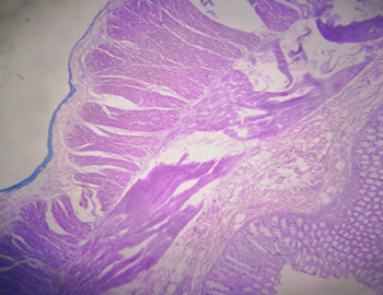
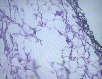
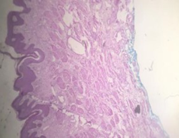
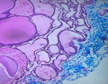
Score: 0-No colour visible; 1- colour is perceptible only faintly; 2- colour is perceptible more clearly than 1 but less than 3, 3-colour perceptibility is clear and sharp The results of black and blue colour were comparable to that of India ink in all types of tissue; in fact poster colours were sharper and easily visible in breast and colon specimen than India ink (all sections of blue and black poster colour gave score 3 visibility as compared to India ink in which only 4/9 (45%) cases of breast and 2/5 (40%) cases of colon gave score 3 visibility) Blue colour yielded the most consistent results in all types of tissues and the results of green colour were poor. Figure 1 shows the visibility of different poster colours in different tissues.
 |
 |
|---|---|
 |
 |

In the present study, the poster colours successfully
marked the tissue margins of routine
histopathology specimens and yielded results
comparable to that of similar tissues marked with
India ink. India ink is the most well known tissue
marking dye. Kizhakkoottu S (2021) 7 conducted a
large scale study on 1325 specimens to compare
the properties of India ink,
Acrylic colours and Tissue Marking Dyes in inking of
tumour resection margins. They concluded that
India ink is the best tissue marking dye but the
advantages of acrylic paints compete with that of
India ink and require much larger studies to prove
their reproducibility. Our study also yielded similar
results with additional benefit of less cost and
multiple colours.
Poster colour is a type of acrylic paint and it is
commonly used in artwork and painting. It is a
distemper paint which has gum water as its binder
and has water soluble characteristics; while acrylic
paints are particulate pigments suspended in acrylic
polymer emulsion which along with a mixture of
multiple chemicals forms a thick emulsion. Poster
paints can be used in the same way as acrylic
paints; except poster paints are more economical
and dries more quickly than regular acrylic paints.
Acrylic paints were first introduced around 1950,
and have become increasingly popular with
multiple uses, as these paints are easy to apply, dry
and are long lasting. 8
Aziz A (2019) & Pursnani D (2015) used acrylic
colours on breast and colon specimen and
concluded that use of acrylic colours has many
benefits over India ink. They also studied three
different methods of colour application and found
that best results were achieved when the sections
were taken after inking and overnight fixation. 6,9
Tampi C. (2012) concluded that the easy availability
and application of acrylic colours mark them as a
good alternate inking technique in India that gives
reliable results on microscopic examination and use
of coloured inks provides precision to the
examination of the tissue margins. 10 Sarode SC et al
(2015) used acrylic paint for margin inking in oral
squamous cell carcinoma and concluded that acrylic
colours are better than India ink as surgical ink
because they are available in a variety of colours,
are easy to apply, quickly dry, and withstand the
processing procedures. They also have an
advantage of being easily available and of being
clearly visible on paraffin blocks and under
microscope. They further suggested use of multiple
colours will aid in reassessment of resection
margins of the gross specimens in the future. 11 We
also experienced similar advantages as the studies
mentioned though our sample size was much less.
Vatsyayan A (2019) 12 , Criswell (2021) 13 , Mardiana AA
(2019) 14 are of the same view regarding the
usefulness of poster colours in marking of surgical
specimen. Mardiana AA (2019) conducted his study
in Malaysia and used poster colours which is a
cheaper version of acrylic paints and found it to be
a reliable alternate to India ink and strongly
recommended its use in routine histopathology.
Many studies are available that showed the
benefits of acrylic paints for margin inking but only
a few; as the present study, are about the
application of poster paints for this purpose. We
favour poster paints as their less cost and a good
comparable yield makes them more suitable for
long term use. However, Tampi C. (2012) showed
that not all shades of acrylic colours suffice as
grossing ink because of their poor visibility in slides
as happened in the present study, the green colour
was not visible on colonic serosa and testicular
biopsy.10
Much larger studies are needed, especially in
tissues requiring decalcification to assess the
reliability of poster paints as tissue markers in
routine histopathology, as is pointed out by
Williams AS et al (2014) 15 and Keifer S (2021) 16 in
their research.
Jennifer S (2019) 17 and Kamat M (2019) 18 studied the
importance of resection margin in oral squamous
cell cancer. They recommended the use of acrylic
paints as substitute for india ink and tissue marking
dyes. According to them, coloured ink is more
useful for better orientation, post grossing
reconstruction of the specimen and reducing the
identification error. The ease of use, quick drying
and easy availability of acrylic paints favours their
use. Fei B (2017) 19 and Hon JD (2019) 20 emphasized
the need of accurate resection margins in oral
cancers. Molecular assessment of resection margins
is a novelty. Among the available tools they
preferred the use of coloured inks for surgical
resection margin assessment.
Local data mentions the use of India ink for
evaluation of surgical resection margins but not the
use of any other multicolour inks like acrylic paints
or poster paints. 21 But there are studies from India,
Singapore and Malaysia which highlight the utility
of coloured inks in surgical specimens.6,9,10
We used poster paints for inking of surgical
resection margins while mostly available data is of
the use of acrylic paints. The advantages of using
poster colours in this study were that black poster
colour was more reliable than India ink in case of
colonic mucosa, there was minimal seepage of
colour into the underlying tissue and margin
staining with poster colours did not affect the
quality of tissue sections or staining when
compared with other non-inked tissues processed
on the same day. The limitations of the study are it
is a single centred study with a small sample size.
The use of poster colours as a routine technique
requires a much larger study with a greater sample
size.
Poster colours are available in most stationery shops. They seem to be a reliable tool for tissue marking when India ink is not available. Their availability in a variety of colours provides them an edge over India ink. To ensure reproducibility of the results these alternative paints should be tested in individual laboratories before applying in routine use.
- Shah K. Postoperative pathologic assessment of surgical margins in oral cancer: A contemporary review. J Oral Maxillofac Pathol. 2018; 22(1):78- 85. doi: 10.4103/jomfp.JOMFP_185_16.
- Sarode G , Sachin C , Pankaj S and Shankargouda P. Histopathological assessment of surgical margins of oral carcinomas and related shrinkage of tumour. Oral Oncol. 2017;2(1):1-4. DOI: 10.1177/2057178X17708078
- Hon D, Chen W, Minerowicz C. Analysis and Comparison of Tissue-Marking Dye Detection via Light Microscopy, Telemicroscopy, and Virtual Microscopy. Am J Clin Pathol. 2019; 151(1): 95–9. DOI: 10.1093/ajcp/aqy117
- Alturkistani A, Tashkandi M, Mohammedsaleh M. Histological Stains: A Literature Review and Case Study. Glob J Health Sci. 2015;8(3):72-79. DOI: 10.5539/gjhs.v8n3p72.
- Aziz A, Bakar S, Kornain N. Poster colours: Pocket-friendly alternative to tissue marking dyes. Turk Patoloji Derg. 2019;35(1):170–171. DOI: 10.5146/tjpath.2017.01394
- Kizhakkoottu S, Jayaraj G, Sherlin HJ, Don KR, Santhanam A. Inking of gross specimens: a systematic review. Arkh Patol. 2021;83(1):49-52. DOI: 10.17116/patol20218301149
- Khramtsov AI, Khramtsova GF, Asaulenko ZP. Application of tissue-marking dyes for pathologic examination of surgical and autopsy specimens. Arkh Patol. 2019; 81(1):40-45. Russian. doi: 10.17116/patol20198101140. PMID: 30830104.
- Pursnani D, Arora S, Katyayani P, Ambica C, Yelikar B. Inking in surgical pathology: does the method matter? A procedural analysis of a spectrum of colours. Turk J Pathol. 2016; 32(2):112-8. DOI: 10.5146/tjpath.2015.01351
- Tampi C. In search of the rainbow: Colored inks in surgical pathology. Indian J Pathol Microbiol. 2012; 55(1):154-7. DOI: 10.4103/0377- 4929.97843
- 11. Sarode C, Sarode S, Patil S. Comparative Study of Acrylic Color and India Ink for their use as a surgical margin inks in oral squamous cell carcinoma. World J Dent. 2015; 6(1):26-30. DOI: 10.5005/jp-journals-10015-1308. Vatsyayan A, Patel P, Mandlik . Surgery Is an Art, Live Yours in Colors: Specimen Color Coding in Head and Neck Oncology. Indian J Surg Oncol. 2019; 10(4):618-619. doi: 10.1007/s13193-019- 00941-6. Epub 2019 May 28. PMID: 31866729; PMCID: PMC6895368.
- Criswell S, Sutton J. Application of dyes to cytology cell blocks and biopsy tissues before processing enhances specimen visualization during embedding and microtomy. Histotech.2021. DOI: 10.1080/01478885.2021.1909357.
- Mardiana A, Bakar S, Kaslina N. Poster Colours: Pocket-Friendly Alternative to Tissue Marking Dyes. Turk Patoloji Derg. 2019; 35(2): 170-71. DOI: 10.5146/tjpath.2017.01394. Williams S, Dakin Hache K. Variable fidelity of tissue-marking dyes in surgical pathology. Histopathol. 2014; 64(3):896–900. doi: 10.1111/his.12328.
- Kiefer S, Huber J, Fullgraf H, Sorensen H, Csanadi A, Stillger N, et al. Alteration of Tissue Marking Dyes Depends on Used Chromogen during Immunohistochemistry.CELLS.2021;10(1):83542. doi:10.3390/cells1004083
- Hon JD, Chen W, Minerowicz C, Thomas S, Barnard N, Gilbert N,et al. Analysis and Comparison of Tissue-Marking Dye Detection via Light Microscopy, Telemicroscopy, and Virtual Microscopy. Am J Clin Pathol. 2019 Jan 1;151(1):95-99. https://doi.org/10.1093/ajcp/aqy117.
- Sattar, AK, Ali B, Masroor I. Feasibility of preoperative tattooing of percutaneously biopsied axillary lymph node: an experimental pilot study. Pilot Feasibility Stud 6, 140 (2020). https://doi.org/10.1186/s40814-020-00682-2

An Official Publication of
Islamabad Medical & Dental College
Volume 11 Issue 3
Editorial
Address of Correspondence
Anum Usman
Email:
dranumusman@gmail.com
Cite this article.Sarwar A, Tanwani K A, Usman A, Moeed K. Comparative Benefits of Tissue Marking by Poster Ink in Histopathology.J Islamabad Med Dental Coll. 2022; 11(3): 175-181 DOI: https://doi.org/10.35787/jimdc.v11i3.751