Inflammatory Myofibroblastic Tumour of Lung, A Rare Entity
Bilal Umair1, Sohail Saqib2, Rashid Hussain 3, Raheel Khan 4, Farhanullah5
1 Professor and Head of Department, Combined Military Hospital, Rawalpindi, Pakistan.
2,4,5 Assistant Professor, Department of Public Health, National University of Medical Sciences, Islamabad, Pakistan.
3 Consultant Thoracic Surgery, Combined Military Hospital, Rawalpindi, Pakistan.
Inflammatory fibroblastic tumour of lung is a rare mesenchymal tumour usually affecting the children and young adults. It is usually considered as a benign entity however it has high potential for recurrence and local invasion. Exact pathogenesis of tumour is not known. Complete surgical excision is the treatment of choice. Due to high chance of recurrence, patients are usually kept on close follow up. Here we present a case of inflammatory myofibroblastic tumour of lung , who presented with history of pain and shortness of breath.She was diagnosed a case of inflammatory myofibroblastic tumour with the help of CT scan chest and CT Guided biopsy.She underwent right sided pneumonectomy and remained symptom free on follow up. Keywords:Inflammatory, Myofibroblasts, Pneumonectomy, Tumour.
Inflammatory myofibroblastic tumour (IMT) is a rare mesenchymal tumour that usually involves lungs, abdominopelvic organs and maxillofacial structures. They constitute less than 1% of adult lung tumours.1The tumour usually occurs in paediatric population and young adults.2 Inflammatory myofibroblastic tumour is also known as inflammatory pseudotumour or plasma cell granuloma.It is usually classified as a benign tumour.3,4 However, tendency for local invasion or recurrence is present and in few reports, rare metastasis has also been reported5.Pulmonary IMT usually presents as a solitary pulmonary nodule or as a mass on chest radiography.6,7Preop diagnosis is usually difficult to establish and often possible after resection of the tumour. The treatment of choice is complete surgical excision.
Here we present a case of a pulmonary IMT in a 21 year old lady, involving the right lung who underwent successful surgical excision.
A 21 year old, unmarried lady, resident of Rawalpindi presented to our OPD with 6 months history of pain right side of chest and shortness of breath on minimal exertion. There were no associated complains of haemoptysis, cough or weight loss. On examination, her vitals were normal and there were decreased breath sounds on right side of the chest. Her chest X-ray revealed a mass involving right hemithorax. CT chest showed a large mass occupying whole of right hemithorax and compressing right mainstem bronchus.(Fig.1).
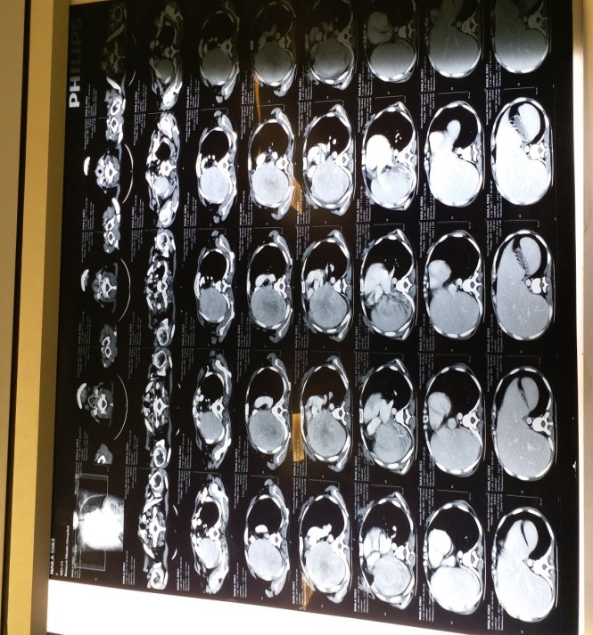
Fig.1 Pre op CT scan Chest
CT guided biopsy of the mass revealed it to be inflammatory myofibroblastic tumour, so surgical excision of the mass was planned after discussion in multidisciplinary meeting including Oncologist,Histopathologist and Radiologist. Patient was counselled regarding treatment plan and prepared for surgery. Standard right sided posterolateral thoracotomy was done and mass was found to be involving whole of the right lung along with involvement of right main stem bronchus, however there was no extension of the mass into the chest wall(Fig.2).Right sided pneumonectomy was performed.
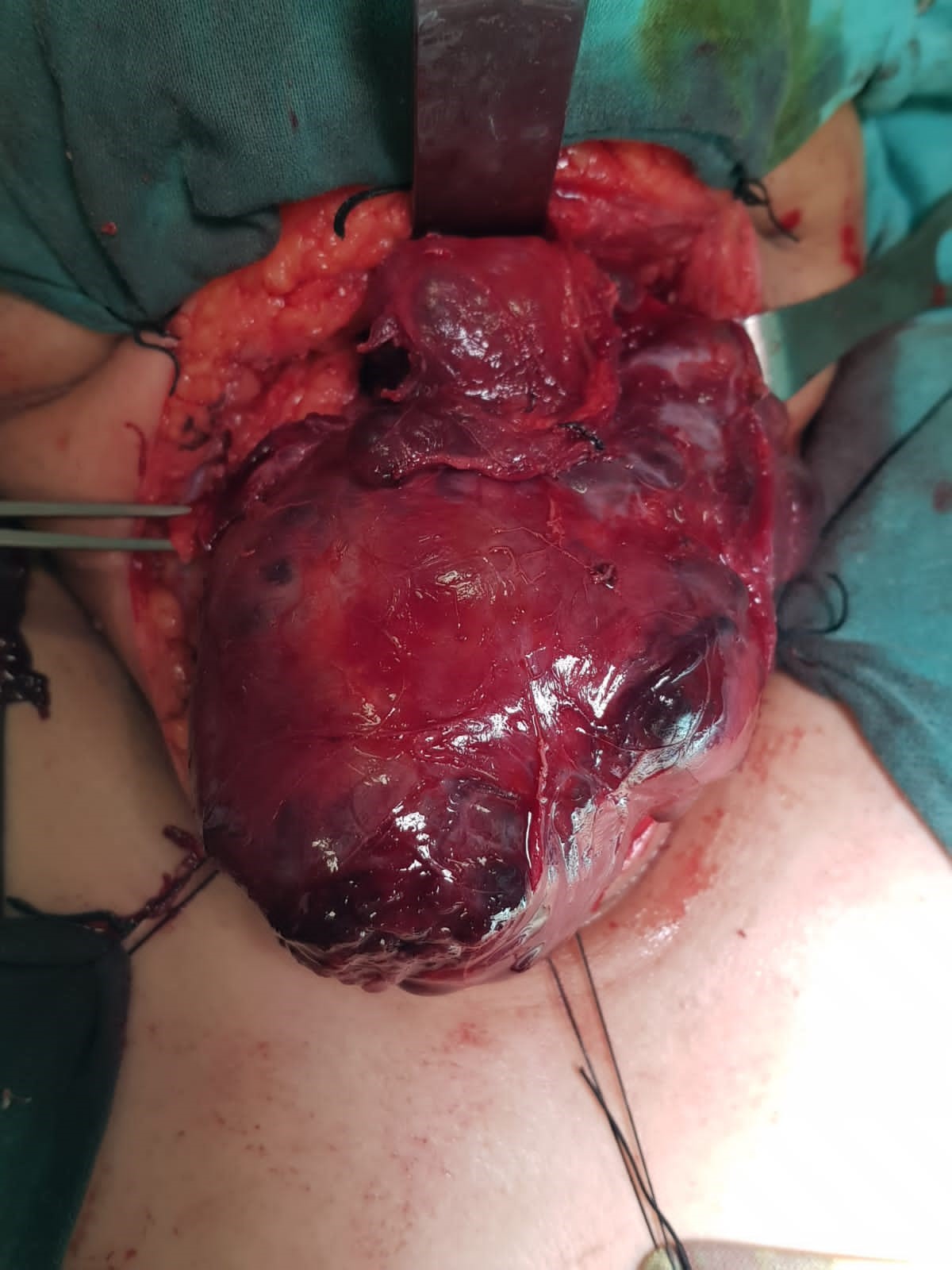
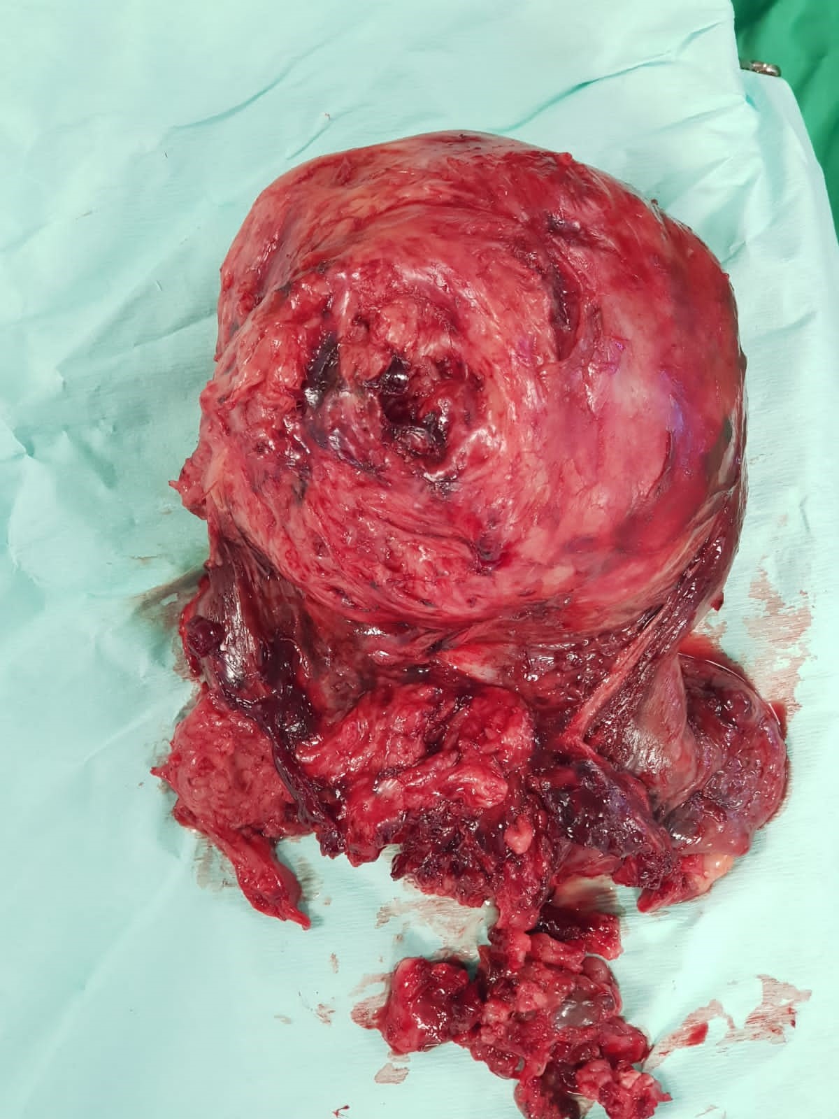
Fig.2 Post operative specimen
Post operatively, patient remained in ICU for 1 day and later on shifted to ward. Post op recovery was uneventful and patient was discharged on 4th post operative day. She was followed up after 1 week with histopathology report. Patient remained asymptomatic in follow up period and histopathology report confirmed the preop diagnosis(Fig.3).She was again discussed in oncology meeting and is placed on close observation and follow up due to high recurrence tendency of the tumour.
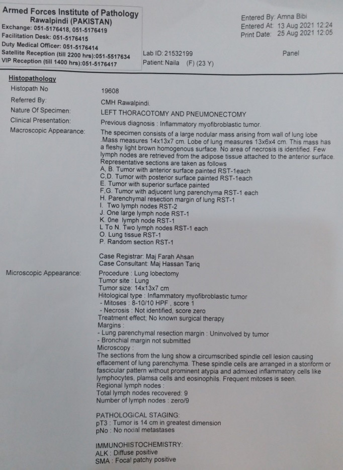
Fig.3 Histopathology Report
Inflammatory fibroblastic tumours of the lung are considered as a rare entity with reported incidence of less than 1 % of all lung tumours in literature.8 IMT is considered as a mesenchymal tumour which is composed of fibroblastic and myofibroblastic spindle cell along with plasma cell,eosinophils and lymphocytes etc. A vast variety of nomenclature has been used in the literature to describe these set of lesions depending upon the predominant cell type. Various names used in literature include plasma cell granuloma, xanthogranuloma, inflammatory myofibroblastic proliferation, inflammatory pseudotumor, fibrous histiocytoma, plasma cell histiocytoma complex and inflammatory fibrosarcoma.9 Matsubara et al used the term inflammatory pseudotumor10 whereas Pettinato et al were among the first ones to use the term IMT due
to the immunohistochemical features of the spindle cells in these lesions resembling those of myofibroblasts.11Exact etiology and pathogenesis are unknown. Though IMT are considered as benign entity, however, malignant transformation has also been reported in the literature including recurrence and rarely metastatic disease. 12
Patients usually present with diversity of symptoms ranging from being asymptomatic and discovered incidentally 13to various symptoms such as cough, chest pain, shortness of breath, hemoptysis, fever and fatigue, with manifestations depending on the
size and location of the tumor.14Our patient presented with symptoms of pain and shortness of breath. Imaging findings are nonspecific and range from benign looking solitary peripheral lung nodules to heterogeneous mass with variable contrast enhancement. When IMT present as central masses, there is often involvement of hilar structures and the mediastinum and so was the finding in our case. Calcification and lymphadenopathy are rare.4 PET can be helpful in distinguishing benign IMT from malignant lesions. Preop diagnosis of IMT is usually difficult and always require histological examination.15Mostly, diagnosis requires surgical excision.
Treatment of choice is complete surgical resection of the tumor. Various therapies such as chemotherapy, radiation and corticosteroids are recommended for patients who have incomplete resection, have multifocal disease, irresectable tumors or when surgery is contraindicated .Prognosis of the disease depends on completeness of surgical excision an size of the tumour. Due to high potential for recurrence of the tumour, patient should be followed up closely even for years to detect local or distant recurrence.
Pulmonary inflammatory fibroblastic tumour,is a rare entity but should always be considered in diagnosis of pulmonary tumours, especially in children and young adults. Despite being considered as a benign entity ,complete surgical exicision and close follow up is required due to high risk of local and rarely distant recurrence.
- A. Al-Obaidi, C. Buess, J. Mogire, P.S. Reddy.Inflammatory myofibroblastic tumor of the lung: an extremely rare condition in adults, Cureus 11 (12) (2019) 6432.
- Coffin CM, Fletcher JA. Inflammatory myofibroblastic tumour. In: World Health Organization Classification of Tumours of Soft Tissue and Bone. 4th edition. Fletcher CDM, Bridge JA, Hogendoorn PCW, Mertens F, editors. Lyon: International Agency for Research on Cancer; 2013. p. 83-4.
- N. Marwah, N. Bhutani, S. Dahiya, R. Sen. Inflammatory pseudotumour:a rare tumor of lung, Ann Med Surg 36 (2018) 106–109.
- Y. Na, S. Park. Inflammatory myofibroblastic tumor of the pleura with adjacent chest wall invasion and metastasis to the kidney: a case report, J. Med. Case Rep. 12 (2018).
- Panagiotopoulos N, Patrini D, Gvinianidze L, Woo WL, Borg E, Lawrence D. Inflammatory myofibroblastic tumour of the lung: a reactive lesion or a true neoplasm?. Journal of thoracic disease. 2015 May;7(5):908-11.
- Yu Y, Wang X, Shi C, Hu S, Zhu H, Hu C. Spectral computed tomography imaging in the differential diagnosis of lung cancer and inflammatory myofibroblastic tumor. Journal of Computer Assisted Tomography. 2019 Mar 1;43(2):338-44.
- Karapolat S, Seyis KN, Ersoz S, Buran A, Karadeniz ZM. Lung image: inflammatory myofibroblastic tumor. Lung. 2017 Jun;195(3):387-8.
- T. Tsuchiya, M. Tanaka. Pulmonary inflammatory myofibroblastic tumor: a case report, Asian Cardiovasc. Thorac. Ann. 26 (2018) 317–319.
- Surabhi VR, Chua S, Patel RP, Takahashi N, Lalwani N, Prasad SR. Inflammatory myofibroblastic tumors: current update. Radiologic Clinics. 2016 May 1;54(3):553-63.
- Matsubara O, Tan-Liu NS, Kenney RM, Mark EJ. Inflammatorym pseudotumors of the lung: progression from organizing pneumonia fibrous histiocytoma or plasma cell granuloma in 32 cases. Hum Pathol 1988;19:807–814.
- Pettinato G, Manivel JC, De Rosa N, Dehner LP. Inflammatory myofibroblastic tumor (plasma cell granuloma). Clinicopathologic study of 20 cases with immunohistochemical and ultrastructural observations. Am JClin Pathol 1990;94:538 –546.
- Sagar AE, Jimenez CA, Shannon VR. Clinical and histopathologic correlates and management strategies for inflammatory myofibroblastic tumor of the lung. A case series and review of the literature. Medical Oncology. 2018 Jul;35(7):1-0.
- Al-Obaidi A, Buess C, Mogire J, Reddy PS. Inflammatory myofibroblastic tumor of the lung: an extremely rare condition in adults. Cureus. 2019 Dec 20;11(12). e6432. doi:10.7759/cureus.6432
- Braham Y, Migaou A, Njima M, Achour A, Saad AB, Mhamed SC, Fahem N, Rouatbi N, Joobeur S. Inflammatory myofibroblastic tumor of the lung: A rare entity. Respiratory Medicine Case Reports. 2020 Jan 1;31:101287.15.
- Sachdev R, Mohapatra I, Goel S, Ahlawat K, Sharma N. Core biopsy diagnosis of alk positive inflammatory myofibroblastic tumor of lung: an interesting case. Turk. J. Pathol. 2018;36:173-7.

An Official Publication of
Islamabad Medical & Dental College
Volume 11 Issue 2
Address of Correspondence
Case Report
Sohail Saqib
Email:
drsohailchatha@hotmail.com
Cite this article.Umair B, Saqib S, Hussain R, Khan R, Farhanullah. Inflammatory Myofibroblastic Tumour of Lung, A Rare Entity. J Islamabad Med Dental Coll. 2022; 11(2):127-131 DOI: https://doi.org/10.35787/jimdc.v11i2.846