Immunohistochemical Expression of Ki-67 in Malignant Salivary Glands Tumors
Sultan Muhammad Wahid1, Faiz Rasul2, Zainab Rizvi2, Khurram Nadeem3, Ahmed Sultan4, Muhammad Talha Haseeb5
1Department of Dental Surgery, PGMI/LGH Lahore, 2Department of Oral Pathology, de’Montmorency College of Dentistry, Lahore; 3Department of Oral Medicine, Lahore Medical & Dental College, Lahore; 4Department of Dentistry, CMH Medical and Dental College, Lahore; 5Department of Internal Medicine, Shaikh Zayed Medical Complex, Lahore
Objective: To determine the expression of Ki-67 in MSGTs to evaluate its role in the diagnosis of salivary gland tumours (SGTs).
Methodology: Sixty cases of MSGTs were collected from the de'Montmorency College of Dentistry, Lahore, from January, 2020 to December, 2021. The histopathological diagnosis and grading of MSGTs was made by H&E staining. On immunohistochemistry Labeling Index (LI) of Ki-67 was determined. Descriptive statistics were calculated on SPSS 22.
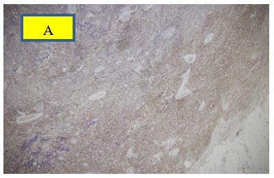
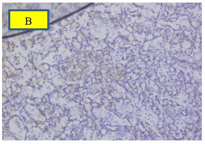
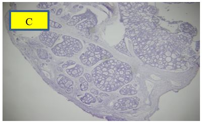
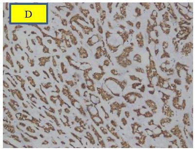
Results: The mean age of the subjects was 51.1 ± 16.70 years. Adenoid Cystic Carcinoma (ADCC) is the commonest type of MSGT, followed by Mucoepidermoid Carcinoma (MEC) and Acinic Cell Carcinoma (ACC), contrary to the worldwide literature where MEC exceeds ADCC. ADCC is mostly located on the palate, but its preferred site, in our setup, comes out to be the parotid gland, followed by the buccal mucosa, and then the palate. In MSGTs, 16.7% were weakly positive, 33.3% were moderately positive, and 50% were strongly positive for Ki-67. There was a very highly significant association (P < 0.0001) between tumour type and Ki-67 LI.
Conclusion: It is concluded that 50% of MSGTs expressed Ki-67 as a strong positive, which is a sensitive indicator of malignancy in salivary gland tumours.
Keywords: malignant salivary gland tumours (MSGTs), mucoepidermoid carcinoma (MEC), acinic cell carcinoma (ACC), adenoid cystic carcinoma (ACC)
Malignant Salivary glands tumors (MSGTs) are extremely rare and they have diverse morphology which creates a great problem in diagnosis.1 World Health Organization (WHO) classified these tumors in more than 30 subtypes.
New classification is simple in which more than 30 subtypes are included.2 MEC is the most common tumor followed by ADCC, ACC and PLGA in MSGTs. Distribution of MSGTs varies according to geographic location and race. Annual incidence of salivary gland tumors (SGTs) is 0.4 to 13.5 cases per hundred thousand cases (100,000) individuals globally. Whereas, out of these cases, malignancies of SGTs are 0.4 to 0.6 per hundred thousand cases (100,000). Proportion of these tumors is 0.3% among all malignancies. Though these tumors have diverse morphology which produces difficulty in diagnosis yet its accuracy play an important role in superior treatment.3 Definitive diagnosis of salivary glands malignancies is based on cell morphology and cellular differentiation. Stromal changes, neural invasion, architecture and growth pattern of the tumor borders are also key features of histological diagnosis, however, still these tumors give diagnostic challenge in certain cases.3
Immunohistochemistry can be used in certain cases where cell type, differentiation, and proliferation are required to assess. Cell proliferation is a basic biological process in tumorogenisis. In certain cases, immunomarkers can be helpful adjunct in the diagnosis of salivary glands malignancies. These immunohistochemical markers can also be used in grading of malignant SGTs.4,5 Among cell proliferative immunomarkers Ki-67 is widely used as a nuclear marker which is a protein in nature. It is detected in all dividing (multiplying/proliferating) cells during active phases of cell cycle except G0. Its antibody (MIB-1) is used to detect malignant cells by routine IHC.
Fundamental principle of this antibody is to detect nuclei of multiplying cells, so it is widely used as adjunct to detect proliferation of cells of malignancies for diagnostic and prognostic purposes.5 Biological behaviour of MSGTs present morphological diversity, and dedifferentiation limit the capabilities of histological assessment, this further enhance in small size biopsies.
A recent analysis of MSGTs revealed highest proliferative index of Ki-67 in ADCC and lowest in PLGA.6 Further there is need to assess Ki-67 proliferative index in different grades of the MSGTs. So, the rationale of this study is to determine proliferative index of Ki-67 in ADCC, MEC, ACC, PLGA, and to determine its usefulness in accurate diagnosis. Another objective is to evaluate its association with various grades of ADCC, MEC, ACC, PLGA such as low grade, intermediate grade and high grade.
Ethical approval of this descriptive study was taken from DCD (de’Montmorency College of Dentistry, Lahore) No. 0261 Dated 13/01/2020. Sample size was calculated by the previous study conducted by Abed et al.7 Samples were collected through non-probability purposive sampling from Department of Oral & Maxillofacial Surgery/Oral Pathology of DCD and PGMI/LGH (Postgraduate Medical Institute, Lahore/Lahore General Hospital Lahore). A total 60 cases of malignant salivary glands tumors such as Adenoid Cystic Carcinoma, Mucoepidermoid Carcinoma, Acinic Cell Carcinoma, and Polymorphous Low-Grade Adenocarcinoma were included in this study. Malignant cases with history of radiotherapy, chemotherapy or recurrence of the tumor were excluded. Clinical data of these patients was taken after consent on the Proforma. Information of the individuals like age, gender, residence, occupation, and site of swelling were taken. Routine laboratory procedures were carried out in Histology/ Oral Pathology laboratory of PGMI, Lahore. Grossing was carried out to examine color, size, and consistency of specimens before proceeding to the laboratory procedures like H&E staining and Ki-67 immunostaining. Principal Investigators confirmed diagnosis of MSGTs. These tumors were graded as Low grade (LG), Intermediate grade (IG) and High grade (HG) according to the study of Rasul et al.3 LG of MSGTs comprised of LG of ADCC (Tubular Pattern), MEC, ACC, and PLGA. IG of MSGTs consisted of Swiss cheese/cribriform pattern of ADCC, and IG of MEC. HG of MSGTs comprised of HG of MEC and HG of ADCC (Solid Pattern).3 Ki-67 cut off value for negative expression was taken as less than 5 malignant cells per 10 HPF (High Power Field). While positive expression was taken as weak positive (WP-1) when 5-19% of malignant cells expressed positivity, moderate positive (MP-2) when 20-49% of malignant cells expressed positivity and strong positive (SP-3) when >50% of malignant cells expressed positivity. Patient record was entered in SPSS version 22. Variables like percentage of cells stained with Ki-67 were described in terms of mean and standard deviation. However, categorical variables such as intensity of Ki-67 were calculated in term of frequencies and percentages. Statistical analysis such as Chi-square test was used to determine the significant association between Ki-67 expression across different types (MEC, ADDCC, ACC and PLGA) and grades (low, intermediate and high) of MSGTs. P value ≤ 0.05 was considered as significant.
Mean age of subjects was 51.1 ± 16.70 years. Adenoid Cystic Carcinoma (ADCC) is the commonest type of MSGT followed by Mucoepidermoid Carcinoma (MEC), and Acinic Cell Carcinoma (ACC) contrary to the worldwide literature where MEC exceeds ADCC. ADCC is mostly located on palate but its preferential site, in our setup, comes out to be parotid gland followed by buccal mucosa and then palate. In MSGTs, 16.7% were weak positive, 33.3% moderately positive and 50% strongly positive for Ki-67. There was a very highly significant association (P< 0.0001) between tumor-type and Ki-67 LI.
Table I: Descriptive Statistics of MSGTs (n=60) |
|||
Variables |
Subgroups |
Frequency |
Percentage |
Age |
Mean ± S.D: 51.1 ± 16.70 years |
|
|
20-40 |
20 |
33.33% |
|
41-60 |
22 |
36.67% |
|
61-80 |
18 |
30.% |
|
Total |
60 |
100 |
|
Gender |
Male |
34 |
56.67% |
Female |
26 |
43.33% |
|
Total |
60 |
100.0% |
|
Site |
Parotid Gland (PG) |
36 |
60% |
Submandibular salivary Gland |
6 |
10% |
|
Sublingual salivary Gland |
0 |
0% |
|
Minor salivary gland on palate |
6 |
10 |
|
Minor salivary gland on tongue |
2 |
3.33% |
|
Minor salivary gland on labial mucosa |
2 |
3.33% |
|
Minor salivary gland on Buccal mucosa |
8 |
13.34% |
|
Total |
60 |
100.0% |
|
Laterality |
Right |
16 |
53.33% |
Left |
14 |
46.67% |
|
Total |
60 |
100.0% |
|
Size |
< 1cm maximum diameter |
4 |
6.67% |
1cm to 2cm maximum diameter |
6 |
10% |
|
2-5cm |
38 |
63.33% |
|
> 5 cm in maximum diameter |
12 |
20% |
|
Total |
60 |
100.0% |
|
Type of Tumor |
MEC |
14 |
23.30% |
ADCC |
32 |
53.33% |
|
ACC |
12 |
20% |
|
PLGA |
2 |
3.33% |
|
Total |
60 |
100% |
|
Grades |
Low Grade |
26 |
43.26% |
Intermediate Grade |
14 |
23.33% |
|
High Grade |
20 |
33.33% |
|
Total |
60 |
100.0% |
|
Invasion |
No invasion (lymphatic & Neural) |
30 |
50% |
Only invasion (Neural) |
28 |
46.6% |
|
Both invasion (lymphovascular & Neural) |
2 |
3.33% |
|
Total |
60 |
100.0% |
|
Ki-67 Expression |
Weak positive |
10 |
16.7% |
Moderate positive |
20 |
33.33% |
|
Strong positive |
30 |
50% |
|
Total |
60 |
100.0% |
|
Table II: Comparison of site and types of Malignant Salivary Gland Tumors (n=60) |
|||||
Site |
Type |
Total |
|||
MEC |
ACC |
ADCC |
PLGA |
||
Parotid gland |
10 |
12 |
14 |
0 |
36 |
Submandibular gland |
4 |
0 |
2 |
0 |
6 |
Palate |
0 |
0 |
6 |
0 |
6 |
Tongue |
0 |
0 |
0 |
2 |
2 |
Labial mucosa |
0 |
0 |
2 |
0 |
2 |
Buccal mucosa |
0 |
0 |
8 |
0 |
8 |
Total |
14 |
12 |
34 |
2 |
60 |
Table III: Comparison of Ki-67 and types of Malignant Salivary Gland Tumors (n=60) |
||||||
Ki-67 Score |
Type |
Total |
p-value |
|||
MEC |
ACC |
ADCC |
PLGA |
|||
Negative Score |
0 |
0 |
0 |
0 |
0 |
≤ 0.0001 |
Weak positive |
8 |
2 |
0 |
0 |
10 |
|
Moderate positive |
6 |
10 |
2 |
2 |
20 |
|
Strong positive |
0 |
0 |
30 |
0 |
30 |
|
Total |
14 |
12 |
32 |
2 |
60 |
|
Table IV: Comparison of Ki-67 findings with Grade of the Malignant Salivary Gland Tumors (n=60) |
||||||
Ki-67 Expression |
Grades of tumors |
Total |
Chi-square |
p-value |
||
Low Grades |
Intermediate Grades |
High Grades |
||||
Weak positive |
6 |
0 |
4 |
10 |
1.226 |
0.268 |
Moderate positive |
14 |
0 |
6 |
20 |
||
Strong positive |
6 |
14 |
10 |
30 |
||
Total |
26 |
14 |
20 |
|
||
100.0% |
100.0% |
100.0% |
100.0% |
|||
MSGTs are more prevalent in Parotid gland followed by buccal mucosa, submandibular gland and palate. MSGTs are also present on buccal and labial mucosa and the tongue. Most of salivary gland tumors are 2-5 Cm in size in our set up. ADCC is the most common type of MSGT in our setup followed by MEC and ACC contrary to the worldwide literature where MEC exceeds ADCC. As per the results of our study ADCC is mostly located in parotid gland followed by buccal mucosa and then palate which is contrary to its worldwide distribution on the palate. Out of 60 MSGTs there was no invasion at all in 30 cases (50%) whereas neural invasion was found in 28 (46.6%) cases and lymphovascular and neural invasion was present in 2 (3.33%) case only. Results show that 50% cases of MSGTs are invasive in nature in our setup. Most of the MSGTs (about 50%) are of low grade and 1/3 (33.33%) are of high-grade malignancy in our setup. High grade malignancies are more common in adenoid cystic carcinoma and mucoepidermoid carcinoma as compared to other two types. When we compared scores of Ki-67 with grades of the MSGTs it revealed that Chi-square test = 1.226 df = 1 and p-value = 0.268 which is not significant in our study. Therefore, it is concluded that ADCC and MEC has high grades in our setup. 50% cases of MSGTs are invasive in nature in our setup. Statistically, there is very highly significant association between type of tumors and Ki-67 scoring (Chi-square test = 47.817, p-value< 0.0001). ADCC has the strongest positive correlation with Ki-67 expression scores followed by moderate scores of ACC and MEC.
Kaza et al., in 2016 determined Ki-67 index in MEC, ADCC, epithelial myoepithelial carcinoma, carcinoma ex PA and adenocarcinoma of SGTs. They also found that Ki-67 was helpful in differentiating low grade and high grade MEC and highlighted malignant behaviour of epithelial myoepithelial carcinoma. They concluded that ki-67 expression is 23% in MSGTs whereas in current study it is 16.7 % weak positive, 33.3% moderate positive and 50 % strong positive. Results of our study are also in congruence with this study where we concluded that Ki-67 is a sensitive indicator in MSGTs.8 Li, in 2015, concluded that Ki67 is highly related to the growth and proliferation of tumor cells. It was also established that Ki-67 outcome is signifi¬cant in malignancies with poorly differentiated cells, as compared to the normal SG tissues. Ki-67 is also a prognostic factor for survival of patients at different stages and phases of the disease. Its antibodies are also used as a treatment with a view to arrest proliferation by micro-injection or oligonucleotides. It is also concluded in our study that Ki-67 is stoutly related to tumor cell proliferation and development because Ki-67 is signifi¬cantly higher in MSGTs.9
Hence the results are in congruence to our study. Faur et al in 2015 determined high expression of Ki-67 in MSGTS which also indicates the usefulness of Ki-67 in identifying highly proliferative forms of salivary gland tumors.10 Sangeetha et al., also reported 17% Ki-67 in MSGTs which is in complete harmony with our results.11 Siddique et al., in 2015 correlated Ki-67 with the grades of MEC. The Ki-67 was low in low grade MEC. Whereas in intermediate grades it was 5-40% with a mean of 25% and in high grades it was 30-75%. Results revealed elevated LI of Ki-67 with higher grade of the MEC. Our results are also same in which LI showed increased values as the grades of the tumors increased.12 In current study most of ADCC (30 out of 32) expressed strong positive expression of Ki-67, only 2 cases out of 32 were moderate positive whereas no case of weak positive was observed. In another study expression of Ki-67 was more expressed in ADCC than MEC similar to current study.13 Similarly, Ki-67 expression was observed more in ADCC as compared to MEC.14 Ki-67 expression was determined in 20 cases of ADCC in which 17 cases (85 %) expressed its expression while 3 cases (15%) were negative however in current study all cases expressed. In current study < 5 % cells were counted as negative while 10 % in Abulhussain’s study.15 In current study no case expressed weak positive staining, only 2 cases expressed moderate positive while 30 cases (93.75%) out of 32 expressed strong positive expression. ki-67 expression was strong positive in high grade of MSGTs.16 In another study, 76.9% ADCC expressed low expression of Ki 67 and only 23.1% expressed as high expression, however in that study low expression was categorized as 11 to 50% positive stain which is moderate positive according to current study. 17 ADCC expressed Ki 67 in all grades but with different range i.e. 27.12 % in grade I, 34.43 % in grade II, and 38.45% in grade III, similarly, in current study it expressed strong positive in grade III.18
In another study Ki -67 expression was higher (40 %) in MSGTs which also support results of the current study in which high expression was observed in MSGTs.19 Ki-67 expressed positive in 96% (24 cases out of 25 cases) in ADCC while it expressed 92% in Polymorphous Low Grade Adenocarcinoma (23 cases out of 25 cases). This is also supporting result of current study in which all cases expressed positive expression in ADCC and PLGA.7 MSGTs have high expression of Ki-67 among them ADCC has highest expression followed by MEC and PLGA similar to the current study.6 High grades MSGTs consistently expressed high expression of Ki-67, however in current study 50% high grade expressed strong positive while 30% expressed moderate positive and 20 % expressed weak positive.20 This is also augmenting that high grade stained strong positive expression of Ki-67 in malignant salivary gland tumors. Raja et al., reported that Mucoepidermoid expressed high expression of Ki-67 as compared to Adenoid Cystic Carcinoma and among different patterns of ADCC tubular pattern expressed high expression than cribriform and solid.21 In another study, Ki-67 expression was highest in malignant salivary gland tumors compared to benign tumors and normal tissues.22 Adenoid cystic carcinomas showed higher Ki-67 expression compared to mucoepidermoid carcinomas and polymorphous adenocarcinomas. It may aid in distinguishing ADCC from other malignant tumors. 23 In another study expression of Ki-67 was highest in ADCC (22.58±0.48) as compared to MEC (21.53±1.52).24 There are certain limitations of the study such as limited cases of MSGTS, and few grades of intermediate grades and high grades. Furthermore, cases of Low-grade polymorphous adenocarcinoma and acinic cell carcinoma were also limited not proportional to mucoepidermoid carcinoma and adenoid cystic carcinoma. Intra oral tumors represent more aggressive behavior as compared to tumors of the major salivary glands, so proportion of minor oral salivary gland tumor is less as compared to parotid tumors which are maximum in the current study. Lastly, literature also showing a variation of scoring system of the Ki-67 regarding categorization of positive cells into weak positive, intermediate positive and strong positive, in current study strong positive score was taken when more than 50% cell stained positive while in literature strong positive score is taken even at less than 30 positive stained cells.
Most cases of ADCC expressed strong positive expression of Ki-67. On the other hand, most cases of ACC expressed moderate expression of Ki-67. While the majority of MEC expressed weak positive expression of Ki-67. Ki-67 expressed as strong positive in most of the cases which indicates its potential role in the development of MSGTs. It may play role in diagnosis of these tumors particularly in small biopsies. High grades also expressed strong positive expression which shows its usefulness in predicting the behavior of the tumor.
Figure A: Ki-67 Immunostaining of high grade Mucoepidermoid carcinoma (X100)
Figure B: Ki-67 immunostaining of Acinic Cell Carcinoma positive expression (X100).
Figure C: Ki-67 immunostaining strong positive expression in ADCC (X100)
Figure D: Ki-67 immunostaining of low grade ADCC showing strong positive expression in (X100).
- Speight PM, Barrett AW. Salivary gland tumours: diagnostic challenges and an update on the latest WHO classification. Diagn. Histopathol. 2020 Apr 1;26(4):147-58. https://doi.org/10.1016/j.mpdhp.2020.01.001
- El-Naggar AK, Chan JK, Grandis JR, Takata T, Slootweg PJ. World Health Organization classification of head and neck tumours. World Health Organization Classification of Tumours. 2017;9.
- Rasul F, Wahid SM, Imran I, Rizvi Z, Jaffar R, Anwar A. Immunohistochemical Expression of BCL-2 in Malignant Salivary Gland Tumors. J Islamabad Med Dent College 2021 Mar 31;10(1):329-36. https://doi.org/10.35787/jimdc.v10i1.542
- Khurram SA, Barrett AW, Speight PM. Diagnostic difficulties in lesions of the minor salivary glands. Diagn. Histopathol. 2017 Jun 1;23(6):250-9. https://doi.org/10.1016/j.mpdhp.2017.04.008
- Bassyoni, Omneya Youssef, Samah Said Elbasateeny, Mahmoud Abdou Yassin, And Taha Abdelwahab Baiomy. Immunohistochemical Expression of Tumour-Associated Macrophage Related Marker (CD68) and Proliferative Marker Ki-67 in Malignant Salivary Glands Tumour: Correlation with the Clinicopathological Factors, Oestrogen Receptor-α and HER-2. J. Clin. Diagnostic Res. 2019. 13(10):10-6. https://doi.org/10.7860/JCDR/2019/42392.13216
- Živković N, Kostić M, Cvetanović A, Milentijević MJ, Spasić M, Petrović M, Trajković M, Denčić T, Krtinić D. Differential challenges in salivary gland neoplasms. Arch. Biol. Sci. 2021 Mar 19;73(1):65-72. https://doi.org/10.2298/ABS201201002Z
- Abed MI, Al-Azzawi LM. Comparative Study of CD105 and Ki-67 in Adenoid Cystic Carcinoma and Polymorphous Low Grade Adenocarcinoma of the Salivary Glands. J. Univ. Babylon eng. sci. 2019 Jun 1;27(3):1-9.
- Kaza, S., Rao, T. J. M., Mikkilineni, A., Ratnam, G. V., and Rao, D. R. 2016. Ki-67 index in salivary gland neoplasm. Int. J. Phonosurg. Larngl., 6(1)-7. https://doi.org/ 10.5005/jp-journals-10023-1110
- Li, L. T., Jiang, G., Chen, Q. and Zheng, J. N. Ki67 is a promising molecular target in the diagnosis of cancer (Review), Mol. Med. Rep. 2015 11(1): 1566-1572. https://doi.org/10.3892/mmr.2014.2914
- Faur AC, Sas I, Motoc AG, Cornianu M, Zamfir CL, Lazăr DC, Folescu R. Ki-67 and p53 immunostaining assessment of proliferative activity in salivary tumors. Rom J Morphol Embryol. 2015 Jan 1;56(4):1429-39.
- Sangeetha, N., Palaniappan, V., Hemavathy, N., and Subathra, K. 2015. Histo- pathological Analysis of Salivary Gland Lesions with Ki-67 immunoprofile. Res. J. Pharma. Biol. Chem. Sci. 2015 5(1): 993-1004.
- Siddique, S., Din, H. U., Hashmi, S. N., Nazar, M., Atique ur Rehman, A. U., Khadim, T., and Akhtar, F. 2015. Role of Ki-67 in the Grading Mucoepidermoid Carcinomas. J. Rawalpindi Med. Coll. 2015. 19(2): 120-123.
- Salehizalani S, Atarbashi-Moghadam S, Mokhtari S, Yazdani F. Expression of Ki-67, P63, P40, and alpha-smooth muscle actin in salivary gland carcinomas with or without myoepithelial differentiation. J. Med. Sci. 2022 Sep 1;42(5):206. https://doi.org/10.4103/jmedsci.jmedsci_204_21
- van der Wal JE, Sgaramella N, Norberg Spaak L, Zborayova K, Nylander K. High podoplanin and low E‐cadherin levels correlate with better prognosis in adenoid cystic carcinoma. Clin. Exp. Dent Res. 2019 Aug;5(4):350-5. https://doi.org/10.1002/cre2.189
- Abdulhussain MM. The Immunohistochemical Expression Ki-67 In Adenoid Cystic Carcinoma of The Salivary Glands. J. Dent. Treat Oral Care. 2018;3(1): 2-5
- Skalova A, Leivo I, Hellquist H, Agaimy A, Simpson RH, Stenman G, Vander Poorten V, Bishop JA, Franchi A, Hernandez-Prera JC, Slouka D. High-grade Transformation/Dedifferentiation in Salivary Gland Carcinomas: Occurrence Across Subtypes and Clinical Significance. Adv. Anat. Pathol. 2021 May 16;28(3):107-18. https://doi.org/10.1097/PAP.0000000000000298
- Chen WL, Wang Y, Zhou B, Liao JK, Chen R. Radical resection and reconstruction in patients with adenoid cystic carcinoma in the minor salivary glands of the palate. Head Face Med. 2022 Dec;18(1):1-9. https://doi.org/10.1186/s13005-022-00312-7
- Bussari S, Jeergal PA, Sarode M, Namazi NA, Kulkarni PG, Deshmukh A, Kulkarni D. Evaluation of Proliferative Marker Ki-67 in Adenoid Cystic Carcinoma: A Retrospective Study. J. Contemp. Dent. Pract. 2019 Feb 1;20(2):211-5.
- Bagulkar BB, Gawande M, Chaudhary M, Gadbail AR, Patil S, Bagulkar S. XIAP and Ki-67: a correlation between antiapoptotic and proliferative marker expression in benign and malignant tumours of salivary gland: an immunohistochemical study. Journal of clinical and diagnostic research: JCDR. 2015 Feb;9(2):EC01
- Skalova A, Leivo I, Hellquist H, Agaimy A, Simpson RH, Stenman G, Vander Poorten V, Bishop JA, Franchi A, Hernandez-Prera JC, Slouka D. High-grade transformation/dedifferentiation in salivary gland carcinomas: occurrence across subtypes and clinical significance. Adv. Anat. Pathol. 2021 May 16;28(3):107-18. https://doi.org/10.1097/PAP.0000000000000298
- Raja R, Shetty DC, Juneja S, Tandon A, Gulati N. MCM3 proliferative index is worthier over Ki-67 in the characterization of salivary gland tumors. Indian J Pathol Microbiol. 2021 Jan 1;64(1):22.
- Doaa, Mansour, Ghoneim., Hanaa, S., Raslan., Omneya, R., Ramadan., Ibrahim, M., Zeitoun. Expression of il-10 and ki-67 in some benign and malignant salivary gland tumors "an immunohistochemical study". Alexandria Dental Journal, (2022). https://doi.org/10.21608/adjalexu.2021.42308.1100
- Saman, Salehizalani., Saede, Atarbashi-Moghadam., Sepideh, Mokhtari., Farzad, Yazdani. Expression of Ki-67, P63, P40, and alpha-smooth muscle actin in salivary gland carcinomas with or without myoepithelial differentiation. Journal of Medical Sciences, (2022).https://doi.org/10.4103/jmedsci.jmedsci_204_2
- Abd, El-Aziz., Enas, Alaa, Eldin. The Association Between Expression of Ki-67 and MMP-7 proteins and Lymph Node Metastasis in Malignant Salivary Gland Tumors. Egyptian dental journal, (2020). https://doi.org/10.21608/EDJ.2020.27386.1099

An Official Publication of
Islamabad Medical & Dental College
Volume 13 Issue 1
Address of Correspondence
Faiz Rasul
Email:
bayfaiz871@gmail.com
Authors’ Contribution: 1,2Conception; Literature research; manuscript design and drafting; 3,4 Critical analysis and manuscript review; 5,6Data analysis; Manuscript Editing.
Cite this article. Wahid SM, Rasul F, Rizvi Z, Nadeem K, Sultan A, Haseeb MT. Immunohistochemical Expression of Ki-67 in Malignant Salivary Gland Tumors. J Islamabad Med Dental Coll. 2024; 13(1): 143-150. DOI: https://doi.org/10.35787/jimdc.v13i1.886.