Endometriotic Stones; Can they exist? A case report
Muneeb Ullah
Assistant Consultant, Department of Surgery, Maroof International Hospital, Islamabad.
Endometriosis is a gynecological pathology that manifests as pelvic pain, infertility and cyclical issues. The presence of endometriotic tissue outside the uterus is the hallmark of endometriosis and commonest sites are ovaries, fallopian tubes, uterosacral ligaments and lateral pelvic peritoneum. Ovarian endometriotic cyst (EC) is typically a chocolate cyst that is often diagnosed by a combination of clinical history, examination and radiological imaging. However, the definitive diagnosis requires histopathological confirmation. The management is complex and requires long term suppression of menstrual cycle and ovarian cystectomy. We hereby illustrate a case of a young female who presented with EC. During a routine daycare laparoscopic cystectomy, a rare finding of endometriotic stones was ascertained per-operatively. Patient remained well post operatively and was discharged in the evening. Keywords: Cystectomy, Cyst, Endometriosis, Laparoscopic
Endometriosis is a chronic gynecological disease
that occurs as a result of endometriotic tissue
outside the uterus. 1 Common sites are ovaries,
fallopian tubes, uterosacral ligaments and lateral
pelvic peritoneum. 2,3
In ovaries, it frequently
presents as endometriotic cyst (EC) that is non-
cancerous and contains chocolate like fluid. 4
EC is
often the underlying culprit responsible for chronic
pelvic pain associated with ovulation and
menstruation in females of reproductive age. 5 The
diagnosis of EC requires a combination of good
history, clinical examination and investigations such
as cancer antigen 125 (CA-125) and ultrasound
imaging. 1 Although the ultrasonography is
generally able to identify EC but definitive diagnosis
is based on laparoscopy and histological
confirmation. 1,6
Differentials while managing such
cysts must include dermoid cyst, hemorrhagic cyst,
neoplasms and uncommonly, abscesses and ectopic
pregnancy. 7
Management of EC is controversial and
depends upon size, symptoms and patient
preferences especially when there are concerns for
fertility. Long term suppression of menstrual cycle
is the mainstay of management for cysts smaller
than 3cm while those greater than 3cm are dealt
with laparoscopic ovarian cystectomy. 8,9 Patients
who have fertility problems are predominantly
planned to proceed for ovarian cystectomy. This
aids in assisted reproductive methods, alleviates
pelvic pain and improves likelihood of successful
oocyte retrieval procedure. 10 By virtue of this
platform, we hereby report a case of a laparoscopic
endometriotic cystectomy which per-operatively
CASE REPORT
revealed endometriotic stones within the cyst
cavity, a rare occurrence. It has not been reported
previously as per our knowledge.
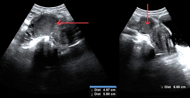
A 30 years old young female, resident of Rawalpindi, presented with cyclical menstrual and pelvic pain in right lower quadrant, dysmenorrhea and irregular cycles for the last two years. Pain aggravated during menstruation and relieved by taking oral non steroidal anti-inflammatory drugs. It was not associated with any physical activity, food, anorexia, weight loss, fever, any symptoms related to gastrointestinal or urinary tract. For the past six months, she was under medical management for endometriosis and right ovarian EC. Recently after her wedding, she and her husband had planned for conception. There were no clinical findings except right sided uterosacral ligament tenderness on pelvic examination. CA-125 levels were slightly raised to 58 U/mL. Recent ultrasound showed right adnexal EC of 5.8 x 4.9 x 6.6 cm in size as depicted by Figure 1.
Due to failure of medical management, chronic pain and fertility related concerns; patient was planned for diagnostic laparoscopy and to proceed for laparoscopic cystectomy. Prior to surgery, informed consent was taken for oophorectomy, cystectomy and deroofing of cyst with possibility of recurrence and insufficient ovarian tissue preservation for fertility. Laparoscopy was performed in trendelenburg’s position. Operative findings were consistent with right EC but additionally a rare finding of endometriotic stones was discovered, shown in Figure 2 and Figure 3.
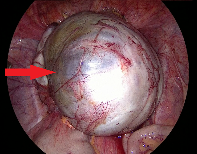
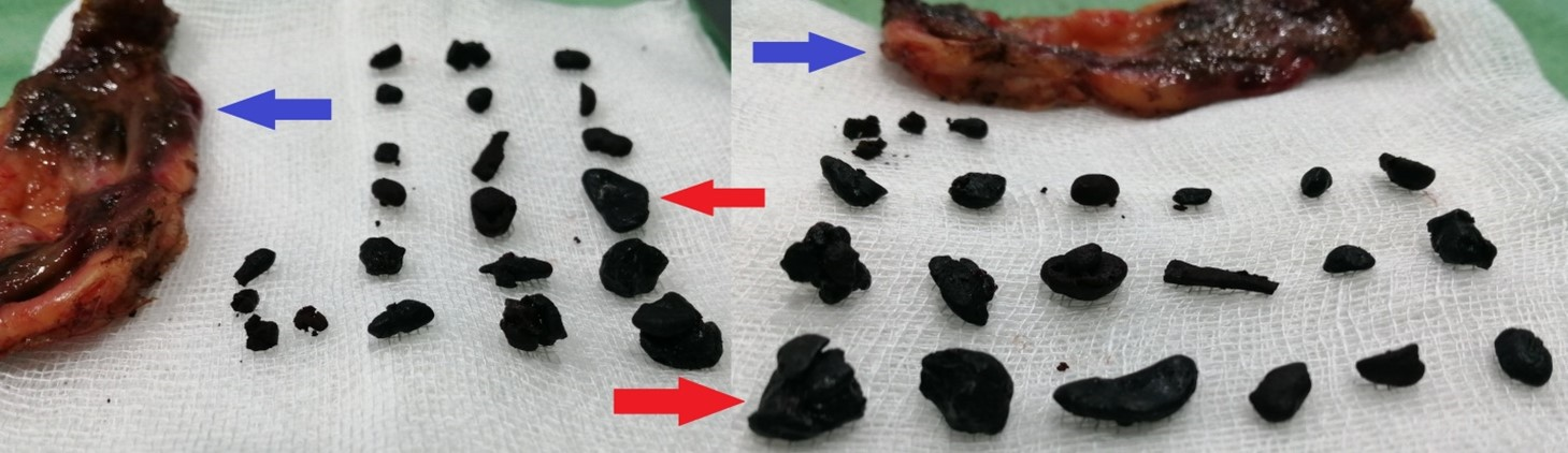
Figure 3: Per-operative image of endometriotic stones (red arrows) Laparoscopic cyst deroofing, followed by cystectomy was performed. Remnant tissue was packed with adrenaline soaked gauze for five minutes. Hemostasis was secured using bipolar electrocautery. Specimen including stones was delivered in bag. Picture of specimens is shown in Figure 4.
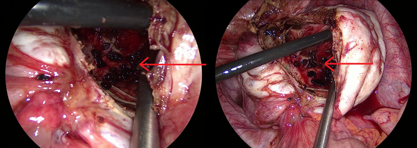
Figure 4: Post operative image showing cyst wall (blue arrows) and endometriotic stones (red arrows) Cyst wall was sent for histopathology. Patient was discharged within 24 hours and had unremarkable post-operative recovery. Histopathology report was consistent with EC.
Endometriosis has variable presentations and
diagnosis is often delayed. This causes hindrance in
proper management and results in vast implications
especially on quality of life and daily routine.11
Laparoscopic ovarian cystectomy is recommended
for a symptomatic EC that is without a widespread
disease and is greater than 3cm size. Although this
provides lowest recurrence rates and highest
possibility of spontaneous conception, the risk of
insufficient ovarian reserve persists. This is
attributed to inadvertent removal of normal
ovarian tissue during cystectomy and cauterization
to control the bleeding. 9 First choice of imaging in
pelvic adnexal pathologies is ultrasonography which
is cheap, effective and without any radiation or
contrast exposure. CT scan and MRI are superior to
ultrasound in assessing the adnexal masses as they
help in correlation of surrounding structures and
surgical planning. 6 Additionally they are able to
differentiate complex cysts, septated lesions, solid
masses and benign from malignant pathologies. We
did not perform a CT scan or MRI since the clinical
diagnosis was quite evident and the size of cyst was
not a huge one. Patient was planned for
laparoscopic intervention which is considered gold
standard for the diagnosis of endometriosis and has
the therapeutic advantage of treating
endometriotic cysts as well. 12 Our patient had
concerns for fertility since she was planning for
conception, and endometriosis at any stage is
known to decrease fertility. 11 Treatment modalities
were discussed with the patient and her husband,
and consensus was reached to proceed surgically.
Laparoscopically, the cyst is usually aspirated under
vision to confirm the diagnosis and to take the
sample. Later, suction and irrigation is performed
multiple times before proceeding for cystectomy to
avoid contamination. In our case, aspiration needle
was used to take the sample as well as for suction
and irrigation, before carrying out cystectomy. The
stones were thus easily seen after deroofing, else
they could have easily been missed. Bleeding from
the ovarian tissue was packed with adrenaline-
soaked gauze, later, hemostatsis was achieved
using bipolar cautery. Monopolar or bipolar cautery
is an effective method for controlling hemostasis
but results in damage to ovarian follicles causing
insufficient ovarian reserve. 13 Adrenaline-soaked
gauze decreases the need for bipolar cautery and
its extent, thus improving outcomes related to
ovarian reserves. Histopathological confirmation is
a requisite for definitive diagnosis and also rules
out any underlying malignancy.
Although laparoscopy has the advantage of speedy
recovery, minimal scar, diagnostic evaluation of the
whole abdominal cavity and staging of the disease,
but it has the disadvantage of decreased depth
perception of the cyst, loss of tactile feedback, risk
of spillage and implantation especially at the port
sites.
Laparoscopic technique has the advantage of being
diagnostic and therapeutic modality. In addition, it
has the capability to stage the disease and identify
uncommon findings that can easily be missed
otherwise. EC can be managed laparoscopically if
the expertise is available. Care must be taken to
avoid port site implantation.
- Foti PV, Farina R, Palmucci S, Vizzini IAA, Libertini N, Coronella M, et al. Endometriosis: clinical features, MR imaging findings and pathologic correlation. Insights Imaging. 2018; 9:149-172. https://doi.org/10.1007/s13244- 017-0591-0
- Katsoulis IE, Katsoulis IE. Retrorectal endometrioid cyst: a case report. J Med Case Reports. 2010; 4:389-10. https://doi.org/10.1186/1752-1947-4-389
- Jiao Y, Lu B. Poorly differentiated mucinous carcinoma with signet ring cells in an ovarian endometriotic cyst: a case report. Diagn Pathol. 2019; 14:73. https://doi.org/10.1186/s13000-019-0850-0
- Hingankar Y, Taksande V and Mahakarkar M. Chocolate Endometrial Cyst: A Case Report. Journal of Pharmaceutical Research International. 2021; 33(40B):1-6. https://doi.org/10.9734/jpri/2021/v33i40B32 254
- Bulun SE, Yilmaz BD, Sison C, Miyazaki K, Bernardi L, Liu S, et al. Endometriosis. Endocr Rev. 2019; 40(4):1048-1079. https://doi.org/10.1210/er.2018-00242
- Yaşar L, Sönmez AS, Zebitay AG, Neslihan G, Yazıcıoğlu HF, Mehmetoğlu G. Huge ovarian endometrioma—a case report. Gynecol Surg. 2010; 7:365-367. https://doi.org/10.1007/s10397-009-0482-6
- Asani L, Nikolova D, Madzunkova K, Karanfilovski V, Pandilov S, Janevska V. Osseous Metaplasia in Ovarian Endometrial Cyst: A case report. 2019; 23(5):17729-17732. http://dx.doi.org/10.26717/BJSTR.2019.23.00 3959
- Vercellini P, Viganò P, Somigliana E, Fedele L. Endometriosis: pathogenesis and treatment. Nat Rev Endocrinol. 2014; 10:261-275. https://doi.org/10.1038/nrendo.2013.255
- Akkaranurakkul P, Lertvikool S, Hongsakorn W, Vallibhakara O, Tantanavipas S, Paiwattananupant K, et al. Effects of intravenous tranexamic acid on ovarian reserve and intra-operative blood loss during laparoscopic cystectomy of endometriotic cyst: a pilot randomized controlled trial. Pilot Feasibility Stud. 2021; 7:171. https://doi.org/10.1186/s40814-021-00907-y
- Dunselman GAJ, Vermeulen N, Becker C, Calhaz-Jorge C, D’Hooghe T, Bie BD, et al. ESHRE guideline: management of women with endometriosis. Human reproduction. 2014; 29(3):400-412.
- Frankel LR. A 10-Year Journey to Diagnosis With Endometriosis: An Autobiographical Case Report. Cureus. 2022; 14(1):21329. https://doi.org/10.7759/cureus.21329
- Young H, Bui T, Cramer SE, O’Connell R, Houshyar R. Ruptured endometrioma in a nonpregnant patient: a case report. J Med Case Reports. 2022; 16:161. https://doi.org/10.1186/s13256-022-03361-3
- Sugita A, Iwase A, Goto M, Nakahara T, Nakamura T, Kondo M, et al. One-year follow- up of serum antimüllerian hormone levels in patients with cystectomy: are different sequential changes due to different mechanisms causing damage to the ovarian reserve?. Fertil Steril. 2013; 100(2):516-22. https://doi.org/10.1016/j.fertnstert.2013.03.0 32

An Official Publication of
Islamabad Medical & Dental College
Volume 11 Issue 3
Editorial
Address of Correspondence
Muneeb Ullah
Email:
muneebullah@gmail.com
Cite this article.Ullah M. Endometriotic Stones; Can they exist? A case report. J Islamabad Med Dental Coll. 2022; 11(3): DOI: https://doi.org/10.35787/jimdc.v11i3.891