Middle Ear Embryonal Rhabdomyosarcoma in a Five Years Old Child A Rare Case Report
Asma Khattak1 , Nosheen Nabi 2, Summaya Sohail Chaudry3
1Medical Officer, Department of Pathology, Pakistan Institute of Medical Sciences, Pakistan
2Assistant Professor, Department of Pathology, Rawal Institute of Health Sciences, Islamabad,, Pakistan
3Assistant Professor, Department of Pathology, Rawal Institute of Medical Sciences, Pakistan
Abstract
Rhabdomyosarcoma is the most common pediatric malignancy with a predilection for head and neck region. Embryonal Rhabdomyosarcoma is a variant of rhabdomyosarcoma which is extremely rare in middle ear. We present a case of middle year embryonal rhabdomyosarcoma in a five-year-old child who was treated as otitis media on first presentation and later misdiagnosed as a vascular aural polyp on histopathology.
Introduction
Rhabdomyosarcoma (RMS) is one of the most frequently occurring malignant soft tissue sarcoma which is thought to be originated from mesenchymal stem cells showing striated muscle differentiation. Based on morphology, RMS is classified into Embryonal RMS (ERMS), alveolar RMS (ARMS) and pleomorphic RMS, in which ARMS and ERMS most commonly occur in children, while pleomorphic RMS is mainly found in adults.1 The most commonly affected areas are head and neck, genitourinary tract, retroperitoneum and extremities. In head and neck orbit is involved in about one-third of the cases while ear is a rare site of involvement. The embryonal RMS comprises about 60%-70% of RMS
cases.2
The diagnosis of RMS of middle ear is usually delayed because it is usually misdiagnosed because of its rare location and its initial symptoms which resemble otitis media and aural polyp .3 It is thus very important to keep RMS in the differentials while dealing with otitis media or aural polyp.
A two years old girl presented in outpatient department with the complain of ear discharge from right ear and was treated as otitis media, she later presented with aural polyp. The biopsy was taken and diagnosis of vascular aural polyp was made on histopathology and she was given treatment for the same. After three years she presented again with a polypoidal growth involving external and middle ear
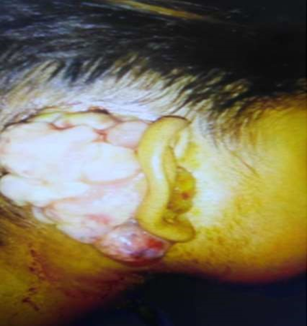
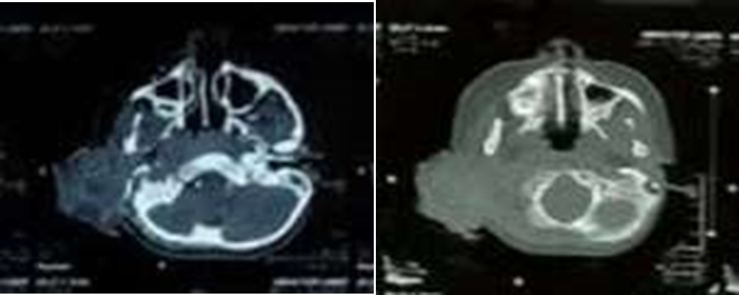
Figure 2: A. Contrast enhanced study showing mass lesion. B. Bone window showing changes in bony part of ear.
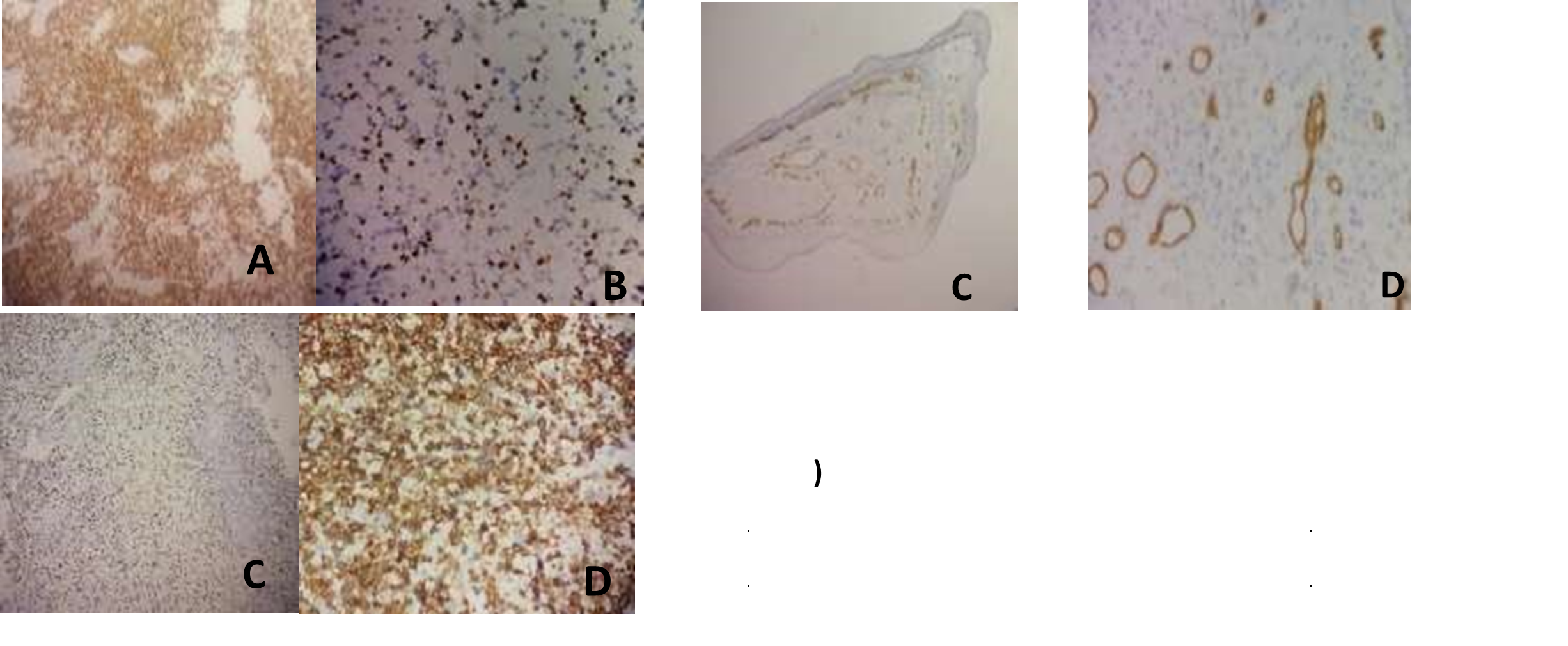
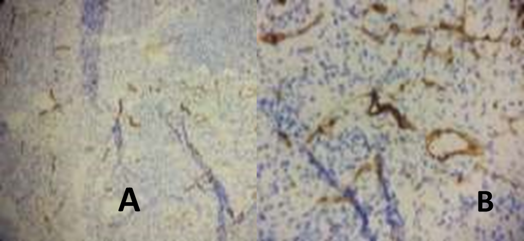
We received two soft tissue fragments in our A laboratory, the largest one measured 2x1.5cm, the entire specimen was submitted. Microscopic cells C: Hypocellular areas, D: Hypercellular areas. Immunohistochemical stains were done which showed strong membranous and nuclear positivity for Desmin and Myogenin respectively, SMA, CD 31, Cd 34 stain was observed only in the endothelial lining of blood vessels, CK 1/3 was seen only in stratified squamous epithelium, LCA was negative (Figure 4) Figure 4: Strong immune positivity was observed for A. Desmin 10x. B. Desmin 40x. C. Myogenin 10x, Myogenin 40x.
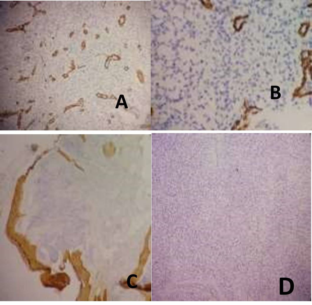
E and F. SMA was seen in vessels only. G. CK 1/3 was positive in stratified squamous epithrlial lining only H. LCA was negative
Rhabdomyosarcoma is an aggressive malignancy and 40 percent cases occur in head and neck region.4 There are three histological types; embryonal, alveolar and pleomorphic, of which embryonal type is most commonly found in head and neck region where its common location is nasopharynx and orbits.5 Very few cases arise from middle ear. Clinical presentation depends upon the structures involved but they usually present as chronic otitis media or aural polyps and treated as such thus delaying diagnosis and leading to advanced disease at diagnosis, when total resection is nearly impossible in most of the cases.6 It is diagnosed with the help of radiological scans and histopathological examination with immunohistochemical stains. MRI usually reveals expansile lytic lesion with low to intermediate signal on T1W and high signal intensity on T2W. Histopathological examination shows round blue cell morphology with positive Desmin and Myogenin stains. The treatment usually is chemoradiotherapy. 7 They have poor prognosis.8 The proximity to the brain and other vital structures makes its poor survival. 9 Patients with stage 1 and stage 2 have good prognosis while intracranial invasion (stage 3) is a factor for poor prognosis.10
Rhabdomyosarcoma of the ear is a rare entity but it should be considered in young children with otitis media or aural polyp as early diagnosis is essential for preventing local spread and metastasis and proper treatment.
- Drabbe C, Benson C, Younger E, Zaidi S, Jones RL, Judson I, et al. Embryonal and Alveolar Rhabdomyosarcoma in Adults: Real-Life Data from a Tertiary Sarcoma Centre. Clin Oncol. 2020 1;32(1):e27-35. doi: 10.1016/j.clon.2019.07.007. Epub 2019 Jul 23
- Ragab AA, Fakoury MM, Kassouma J, Moustafa K, Al Salem FA. Botryoid rhabdomyosarcoma in mastoid and middle ear in a 4-year-old boy: A rare case report. Hamdan Med J. 2018. 1;11(3):130. DOI: 10.4103/HMJ.HMJ_6_18
- Bhandarkar A, Menon A, Kudva R, Pujary K. Embryonal Rhabdomyosarcoma-A Mimicker of Squamosal Otitis Media. Iranian J Otorhinolaryngol. 2020;32(108):57. doi: 10.22038/ijorl.2019.38807.2280
- Attakkil A, Thorawade V, Jagade M, Kar R, Rohe D, Hanowate R, et al. Our Experience with Embryonal Rhabdomyosarcoma Presenting as Aural Polyp. Int J Otolaryngol and Head & Neck Surgery. 2014 Dec 24;4(01):1. DOI: 10.4236/ijohns.2015.41001
- Bhandarkar A, Menon A, Kudva R, Pujary K. Embryonal Rhabdomyosarcoma-A Mimicker of Squamosal Otitis Media. Iranian J Otorhinolaryngol. 2020;32(108):57-61
- Menzies-Wilson R, Wong G, Das P. Case Report: Case report: A rare case of middle-ear Rhabdomyosarcoma in a 4-year-old boy. F1000Research. 2019;8. DOI: 10.12688/f1000research.20558.2
- Beghdad M, Mkhatri A, Berrada O, Abada R, Mahtar M. Embryonal mastoid rhabdomyosarcoma in a three years old child: A case report. Int J Surg Case Reports.

An Official Publication of
Islamabad Medical & Dental College
Volume 12 Issue 1
Address of Correspondence
Nosheen Nabi
Email:
nosheennabi@yahoo.com
Cite this article.Khattak A, Nabi N, Chaudry S S. Middle Ear Embryonal Rhabdomyosarcoma in Funding Source: Nil a Five Years Old Child - A Rare Case Report. J Islamabad Med Dental Coll. 2023; 12(1):57-60 DOI: https://doi.org/10.35787/jimdc.v12i1.923